Meibomian Gland Disease

Earn CME/CE in your profession:


Continuing Education Activity

Meibomian gland disease (MGD) is a broad term involving meibomian gland dysfunction that results in altered meibomian gland secretions and tear-film abnormalities. MGD can be congenital or acquired but is more commonly acquired in nature. It is seen more commonly with advancing age but also typical in youngsters, particularly those involved in excessive digital device use, exposed to wind or extreme sunlight conditions, or contact lens users. MGD affects the meibum secretions, which leads to blocked meibomian ducts and thus decreased lipid secretion, altering the tear film composition. The reduced tear film stability results in increased evaporation, hyperosmolarity, inflammation, and ocular surface disorder. MGD severity is based on staging, including symptoms, corneal and conjunctival staining, and quality of meibum secretions. Diagnosis is based on simple tests that can be done in the outpatient department and includes the determination of ocular symptoms using specific questionnaires. Treatment depends on whether the patient is diagnosed in an asymptomatic or symptomatic stage and severity at treatment initiation. This activity highlights the role of the interprofessional team in the management of patients with meibomian gland disease.

Objectives:

  • Outline the pathogenesis of meibomian gland disease.
  • Summarize the clinical features and staging of meibomian gland disease.
  • Describe the management protocols for meibomian gland disease.
  • Explain the close differential diagnosis for meibomian gland disease.

Introduction

The meibomian gland dysfunction (MGD) international workshop describes MGD as "chronic diffuse abnormality of the meibomian glands, characterized by terminal duct obstruction along with qualitative or quantitative changes in the glandular secretion."[1] The obstruction of the terminal duct occurs because of hyperkeratinization of the duct epithelium and the viscous nature of the meibum. These can result in atrophy and drop out of the meibomian glands, thus decreasing secretion. The sequelae of MGD can be observed in the forms of tear film abnormalities, ocular surface irritation, inflammation, or ocular surface disease.[2] 

These meibomian glands are a type of sebaceous glands that are located in the eyelids. These are named after a German physician and anatomist 'Heinrich Meibom.'[3] These glands lie parallelly in a single row within the tarsal plates of the upper and lower lids. The proximal ends of the meibomian glands extend towards the proximal margin of the tarsal plates. The distal end of the tarsus receives the secretion, i.e., meibum, through the excretory duct into the lid margin. It is estimated that the upper lid contains approximately 20 to 30 separate glands, and the lower lid has about 40 to 50 glands.[4] 

Meibomian glands produce lipids, which are the major component of the superficial lipid layer of the tear film. This lipid layer is known to protect the excessive evaporation of the tear film's aqueous layer. It also helps by lowering the surface tension and thus stabilizing the tear film. It acts as a lubricant during blinking and provides an outer barrier that prevents bacteria from entering the tear film. The functional disorders caused by the meibomian gland dysfunction resulting in alterations in the meibomian gland secretions are also referred to as posterior blepharitis.[5] MGD can broadly be classified based on onset into congenital and neoplastic. Based on the duration, it can be classified into acute or chronic.

Based on underlying etiology, it can be divided into:

  • Low delivery
  • High delivery

The low delivery type can be hyposecretory (meibomian sicca) and obstructive (cicatricial or non-cicatricial). Meibomian sicca results from atrophy of the meibomian glands or secondary to medications. MG atrophy results in an overall reduction in functional meibomian glands. The most common form of MGD is the obstructive type. This results from hypertrophy and keratinization of the ductal epithelium. This is further classified into cicatricial or non-cicatricial.[6] The high delivery type is also called hypersecretory (meibomian seborrhea) and results from excessive secretion of the lipids. Meibomian seborrhea occurs secondary to underlying systemic conditions such as seborrheic or atopic dermatitis and acne rosacea.[7]

Etiology

The underlying risk factors that can lead to MGD include age, androgen deficiency, history of atopy, systemic associations like Sjogren syndrome, Steven-Johnson syndrome, psoriasis, acne rosacea. Ocular predisposing factors include the use of contact lenses, eyelid tattooing, chronic blepharitis, or trachoma.[8] 

Infestation of eyelashes by Demodex folliculorum also affects the functioning of meibomian glands. Systemic use of antibiotics, isotretinoin for acne, antidepressants, or hormone replacement therapy affects meibomian glands' functioning.[9] The cicatricial MGD is associated with trachoma, erythema multiforme, and pemphigoid. Non-cicatricial MGD is often associated with acne rosacea, atopic dermatitis, seborrheic dermatitis, and psoriasis.

Epidemiology

MGD is a widespread disorder prevalent worldwide. A meta-analysis by Hassanzadeh et al. found the estimated pooled global prevalence of MGD 35.8%.[10] They also reported MGD to be more common in men than women. Another study by Alghamdi et al. among the Miami population found that 59% of study participants had at least one abnormal MG parameter, which was more common in the elderly population.[11]

The researchers also found that whites had more frequent eyelid vascularity abnormalities (31% versus 1%, p-value <0.0005). There were no significant inter-racial differences noted in regards to meibum quality. Badian et al., in their study from the United States, studied subjective dry eye patients and found MGD prevalence as high as 93.8% among these patients. Further, they did not report significant differences based on age (p-value 0.302) or gender (p-value 0.079).[12]

Pathophysiology

The obstructive type of MGD is the most common form. The continuous meibum secretion results in increased pressure with the meibomian glands. This increased pressure results in ductal dilatation, duct dropouts, acinar degeneration, and ultimately, meibocytes.[5]

The stagnation of the meibomian secretions results in alterations like increased melting points, loss of clarity, and bacterial colonization. Bacteria, most commonly Staphylococcus epidermidis, Staphylococcus aureus, and Propionibacterium acnes, produce lipolytic enzymes and release inflammatory mediators.[13] The released lipolytic enzymes act on the lipids in the tear film and lead to lipolysis, thus resulting in loss of tear film integrity. The instability of the tear film results in signs and symptoms of evaporative dry eye due to increased evaporation of the aqueous layer and hyperosmolarity.[14] The changes in the tear film's flora also lead to the release of toxins, which result in eyelid inflammation.

Histopathology

Gutgesell et al. conducted histopathological examination among patients with MGD and found obstructed and dilated ducts, enlarged acini with cystic degeneration, increased inflammatory cells, and abnormal keratinization. Additional findings reported were squamous metaplasia and granulation tissue formation.[15]

In another study by Obata et al., similar findings were reported from an autopsy of 83 patients. The changes reported were cystic dilatation of ducts and acini, thickening of basement membrane along with acini atrophy, granuloma formation, and lipogranulomatous inflammation.[16]

History and Physical

MGD patients can present asymptomatically or be severely bothered to the extent that their daily routine activities are affected. The common symptoms resemble an evaporative dry eye and include itching, burning sensation, redness, irritation, soreness of eyes, eyelid edema, foreign body sensation, watering, or blurred vision.[2] The patient might also experience difficulty in using contact lenses.

All the patients presenting to an eye clinic should ideally be examined by gland expression by applying moderate pressure over the lower lid. This will help in detecting many asymptomatic MGD patients. Evaluation among symptomatic patients should include checking the lid morphology, meibomian gland mass, expressibility from the meibomian gland openings, tear film volume, tear film break up time (TBUT), Schirmer's test, fluorescein clearance test, conjunctival and corneal staining.

  • Lid morphology – look for lid margin irregularity, pouting or plugging of the orifices, vascular engorgement, changes in the mucocutaneous junction.[17] Any signs of eyelid thickening, dimpling/notching/pitting, tenderness, ridge formation between meibomian glands, or distichiasis should also be noted.
  • Expressibility from the meibomian gland openings – usually, all gland openings should be patent and clear fluid expressed out of the openings.[18] The number of functional glands, obstructive capping/plugging, the color of meibum, and consistency should be noted.
  • Tear film volume – tear meniscus should be >10 mm[19]
  • Tear film break up time (TBUT) – 15 to 45 seconds normal, 10 to 15 seconds borderline, 10 seconds abnormal
  • Tear osmolarity – hyperosmolar tears are irritative to the ocular surface
  • Schirmer's test – Whatman number 41 strips are used. These are 5 mm wide and 35 mm long. Values of 15 mm are normal, 5 to 10 mm are borderline, and <5 mm are abnormal
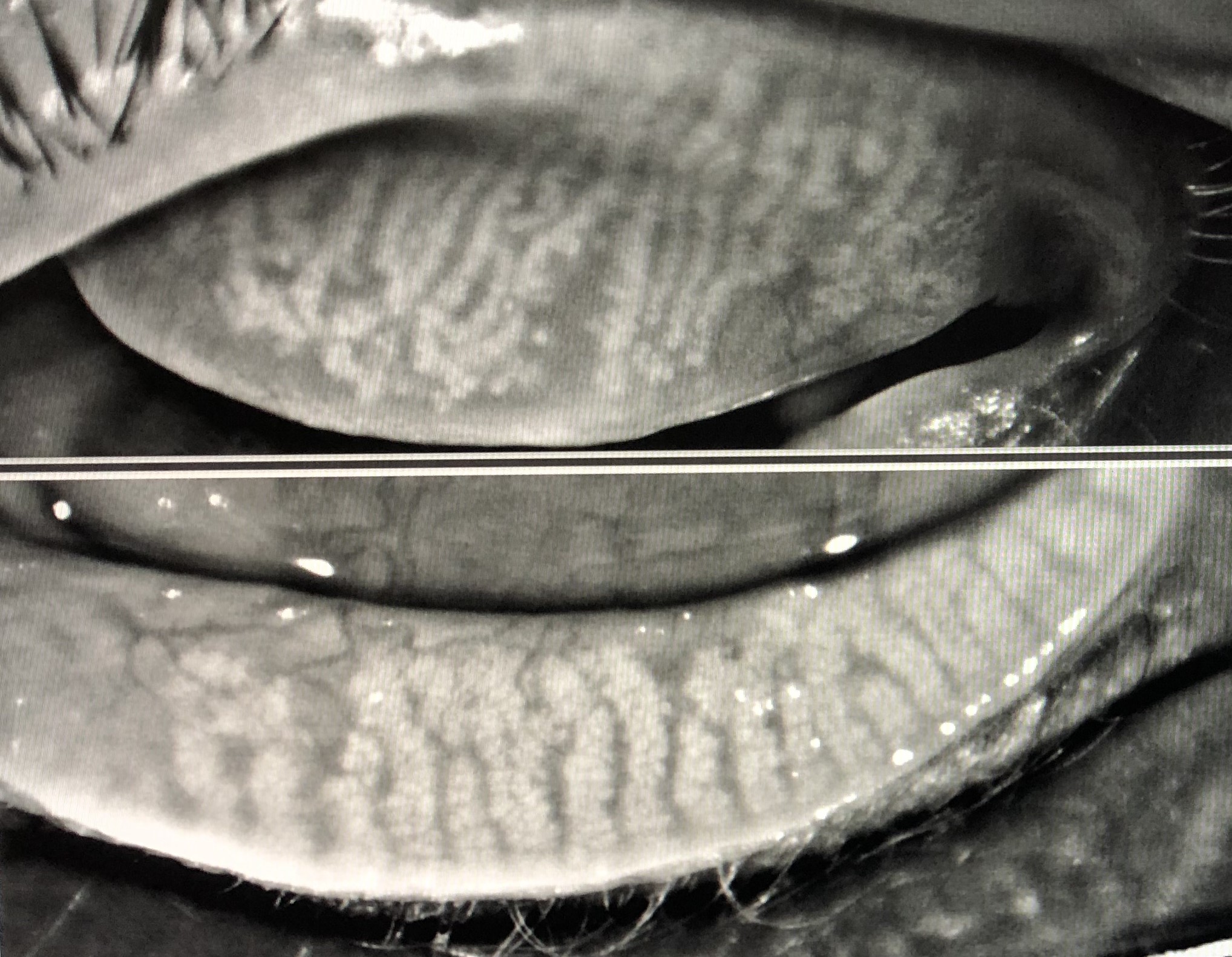
  • Meibography – to check meibomian gland mass, gland loss, shortening, or any irregularities[20] 
  • Fluorescein clearance test detects residual fluorescein after 15 minutes of instilling a drop of 2% fluorescein in the culdesac.
  • Conjunctival and corneal staining – Normally, the conjunctiva and cornea should be clear to Sodium fluoride or lissamine green or rose Bengal staining, with no lid wiper epitheliopathy.

Additional tests that help understand meibomian glands' morphology and functioning include meibometry, meibography, interferometry to check the tear film lipid layer thickness and spread time, and flow cytometry for biomarkers.

Tapie described Meibography in 1977, who used UV woods light to fluoresce the meibomian ducts on biomicroscopy and infrared light to illuminate the meibomian glands.[21] The imaging technique is helpful for direct in vivo visualization of morphology of meibomian glands. The meibography methods have evolved. The methods available include infrared meibography, laser confocal meibography, and optical coherence tomographic meibography. An example of one such system is the LipiView analyzer which uses interferometry (specular reflection) and allows high-resolution images of the lipid layer.[22] The average lipid layer thickness is 60 nanometers. A thin lipid layer is seen in patients with MGD and often corresponds to symptoms.

Results of meibography are measured in the form of the meiboscore or meibograde method. The meiboscore for each eye's upper and lower lids is calculated separately and graded from 0 to 3; thus, a total score out of 6 is given for each eye.[23] The scoring is based on the following points:

  • The lid has partial or missing glands
  • The affected lid area is <33%
  • The affected lid area is 33-66%
  • The affected lid area is >66%

The meibograde method is the most comprehensive scoring system for evaluating meibomian gland morphology. The characteristics noted include gland distortion, gland shortening, and gland dropout.[24] Each characteristic is given a score from 0 to 3, and thus each eyelid is separately given a score from 0 to 9.

Evaluation

The diagnosis of MGD is straightforward based on clinical examination. A detailed assessment is needed to look for associated signs of ocular surface damage or dry eyes. A step-wise approach for evaluating patients presenting with ocular surface disorder symptoms can be planned based on initial clinical examination.

  • Symptom questionnaire – A validated questionnaire is useful in quantitatively measuring symptoms resulting from MGD. Paugh et al. developed MGD specific questionnaire using psychometric approaches among noncontact lens users.[25] Other important questionnaires for symptom assessment include the Ocular Surface Disease Index [OSDI] and the Dry Eye Questionnaire [DEQ][26] 
  • Calculate the blink rate and note the pattern of blinks
  • Measure lower tear meniscus height
  • Measure tear osmolarity
  • Measure tear film break-up time
  • Schirmer's test
  • Grading of conjunctival and corneal staining
  • Mass spectrometry – To look for the presence of inflammatory cell markers, provides information regarding the overall ocular surface inflammatory status 33760148
  • Interferometry – for lipid layer assessment

If the diagnosis is still not precise, the patient should be assessed for:

  1. Quantification of morphologic lid features
  2. Quantification of meibum expressibility and quality
  3. Meibography: quantification of dropout

MGD Staging is Based on Symptoms, Expressibility and Secretion from the Meibomian Glands, and Corneal Staining.[4]

Stage Symptoms Meibomian gland secretions Signs – corneal staining
I None Minimally altered expressibility and secretion quality None
II Mild Mildly altered expressibility and secretion quality None to limited to the periphery
III Moderate Moderately altered expressibility and secretion quality Moderate, mainly peripheral
IV Marked Severely altered expressibility and secretion quality Marked, reaches the central area
Plus disease Co-existing disorders of the ocular surface or eyelids  -  -

Treatment / Management

Treatment for MGD is defined based on the signs and symptoms. The international workshop on MGD described treatment based on the stage of the disease.[27] 

Stage 1 – The patient should be explained about the disease and the effect of diet, home, or work environment on evaporation of tears. The result of medications taken for any other systemic conditions should also be evaluated. Warm compresses and lid expression are usually sufficient in this stage. Warm compresses aim to apply heat to the lid margins directly. The localized increased temperature leads to the melting of the altered meibomian secretions. With regular adherence to the treatment, symptomatic relief is reported by the patients.[28] 

Digital massage of the lids following warm compresses allows expression of the thicker meibum from the glands. This manual expression of altered secretions clears the meibomian gland ducts blockage, allowing the meibomian glands to produce normal secretions. Regular lid hygiene with warm water lid scrubs, with or without local shampoo application, are also recommended to patients.[29] Patients are advised to clean the base of the eyelashes posterior to the lid margin. This helps reduce the bacterial load and clearing the meibum, thus reducing resultant inflammation.

Stage 2 – The patient should be advised to optimize the work environment avoid excessive screen time and direct exposure to wind.[30] This helps by decreasing the rate of tear film evaporation. Regular eyelid hygiene with warm water for a minimum of four minutes, once or twice daily, is advocated. This should be followed by a firm massage to express inspissated secretions. Additionally, dietary omega-three fatty acids consumption should be increased.[31][32] 

Additional treatment options like artificial lubricants, topical antibiotics like azithromycin, the emollient, or liposomal spray should be considered case-to-case basis. Topical 1.0% ophthalmic solution of azithromycin is a broad spectrum, macrolide antibiotic with anti-inflammatory properties. Oil emulsion tear substitutes help improve the lipid layer thickness and thus improve the overall tear film stability.[33] Oral tetracyclines have also shown clinical improvement because of their anti-inflammatory effects.

Stage 3 – All the above treatments should be considered in this stage. Additionally, topical anti-inflammatory drugs should be considered for treatment in this stage.[34][35] Lubricant eye ointment should be used at bedtime for symptomatic relief.

Stage 4 - As symptoms and signs are more severe, additional anti-inflammatory drugs are recommended. Topical anti-inflammatory eye drops like 0.05% cyclosporine A and topical steroids like loteprednol etabonate have shown benefits in reducing inflammation in stage 4 MGD.[35]

Plus disease – Targeted treatment might be needed based on a specific condition associated with MGD.

  • Ocular surface disease – Pulsed dose of steroids
  • Phlyctenular keratitis – Steroids
  • Lash trichiasis – Epilation or cryotherapy
  • Chalazion – Intralesional steroids or incision and curettage
  • Anterior blepharitis – Lid hygiene, warm compresses, topical antibiotic
  • Demodex infestation – Tea tree oil scrubs

Surgical Treatment 

In some patients with severe MGD, if conservative management does not benefit much, alternative measures like the mechanical opening of the terminal ducts and meibum expression play an important role. Probing or electronic heating devices allow the opening of blocked ducts either mechanically or assist in the expression of meibum by heating.

  • Intraductal probing is a slit lamp procedure that involves mechanical opening and dilatation of the blocked meibomian gland orifices and ducts.[36] Maskin probes of 1 to 2 mm are initially used to penetrate the gland orifices, followed by larger size probes of 4 to 6 mm to achieve ductal patency. Probing helps by releasing the meibum, opening the blocked ducts, and allowing better action of topical medications. The treatment shows improvement of TBUT, meibum lipid levels, viscosity, lid margin abnormalities, conjunctival hyperemia, and symptomatic improvement among patients.[37] 
  • Dynamic probing – Syed and Sutula described a modified technique of probing that is done against resistance and has shown benefit in a more significant subset of patients.[38] The method involves holding the lid with forceps and pulling it in a direction opposite to the movement of the probe.
  • Electronic heating devices – These help by locally raising the temperature of eyelids and thus helping in melting of the inspissated meibum.[6] The melting point of meibum increases in MGD because of altered meibomian composition. Increased eyelid temperatures help by decreasing the meibomian lipid viscosity and increasing lipid levels of the tear film.
  • LipiFlow – Several electronic heating devices are now available in the market. LipiFlow (TearScience, Morrisville, NC, USA) has been used widely and proved beneficial. 5098463 This device acts by delivering simultaneous heat to the palpebral conjunctiva of both upper and lower lids. It also helps express the meibomian gland content by providing pressure to the outer eyelid surfaces in a pulsatile manner. LipiFlow treatment has improved meibomian gland secretion, TBUT, increased lipid layer thickness on interferometry.[39] Additional benefits in symptomatic relief from irritation and decreased bulbar congestion have also been reported.[40][41] It is important to look for at least six open meibomian glands in the lower lid. The ideal patient who is expected to benefit can have severe dry eye, but it is vital to have some open glands.[4] 
  • MiBo Thermoflow – MiBo thermoflow (MIBO Medical Group, Dallas, TX, USA) is another model of an electronic heating device.[42] The device uses an external paddle heated to 42.2 degrees C and applied with a gel buffer to the eyelids.
  • Intense pulse laser (IPL) therapy – IPL therapy uses a light source that generates wavelengths in the range of 500 to 1200 nm.[43] When applied through the skin, these waves get absorbed into the blood vessels near the skin surface. These waves are then uptaken by the oxyhemoglobin in the blood cells, which leads to local heat generation and coagulation, resulting in blood vessels thrombosis.[44] As a result, patients also experience decreased redness due to abnormal telangiectatic vessels. Further, IPL also helps in eliminating bacteria from the treated area.
  • Neurostimulation – Recently, the role of intranasal tear neurostimulation has been advocated for the treatment of dry eyes associated with MGD. The technique is based on neural pathways connecting the nasal mucosa to structures that maintain the tear film. The anterior ethmoidal nerve is stimulated with the help of intranasal prongs, which results in increased tear volume, decreased tear osmolarity, and improved lipid and protein concentration.[45] The treatment is recommended daily for 3 minutes a day, and this needs to be repeated for three weeks for optimal results.

Differential Diagnosis

The close differentials that mimic meibomian gland disease include diseases that cause eyelid inflammation, conjunctival congestion, or dry eyes.

  • Staphylococcal anterior blepharitis – This condition is characterized by hard deposits in the eyelashes, loss of eyelashes, and distorted lashes or trichiasis. There is often associated ulceration or notching of the lid margin. Other associated signs include dry eyes, punctate erosions, vascularisation, and infiltrates in the cornea. This is commonly associated with atopic dermatitis.
  • Seborrheic anterior blepharitis – This is a condition characterized by soft deposits, with or without loss or distorted eyelashes. Other signs include dry eyes, punctate erosions, vascularisation, and infiltrates in the cornea. This is associated with seborrheic dermatitis.[7] 
  • Demodex blepharitis – characterized by cylindrical dandruff-like scaling (collarettes) around the base of eyelashes.[46] The mites can be seen under high slit lamp magnification.
  • Evaporative dry eyes - present with irritation, burning or stinging sensation, fatigue, blurred vision, intolerance to bright light, and contact lenses. On examination, Schirmer's and TBUT scores are decreased along with decreased tear film height. Ocular study reveals corneal punctate epithelial erosions, positive conjunctival and corneal staining. Severe dry eyes can be associated with autoimmune diseases like Sjogren's syndrome, rheumatoid arthritis, systemic lupus erythematosus, or diabetes.[47] 
  • Conjunctivitis – presents with redness, watering, lid edema, intolerance to light. Signs include diffuse congestion, follicles, papillae, matted eyelashes with or without corneal punctate epithelial erosions.
  • Cicatricial conjunctivitis – Stevens-Johnson syndrome, mucous membrane pemphigoid.[48] 
  • Contact lens-related keratoconjunctivitis
  • Keratitis – neurotrophic, filamentary, interstitial, contact lens-related

Prognosis

The prognosis depends on the stage of the disease and compliance of the patient towards the advised treatment. Simple practices like daily eyelid hygiene, keeping surroundings humidified, good hydration, avoiding smoking, regular lubrication, frequent and regular blinking, and sunglasses to protect eyes from direct wind or sunlight are practical ways to prevent worsening of MGD and associated evaporative dry eyes symptoms. Early stages of MGD can be managed with topical medications alone and can be reversed with good compliance from patients.[35]

Advanced stages might need probing, thermal heating devices, or pulsed laser therapy. Severe dry eyes might lead to blurred vision, inability to wear contact lenses, headaches, corneal keratinization, scarring, or corneal ulcers. Thus, it is imperative to treat MGD patients effectively and follow up to prevent profound vision-threatening complications.

Complications

MGD is seen as a mild disease by many ophthalmologists and is often overlooked in the early stages. MGD, if ignored and not treated correctly in the early stages, can lead to severe symptoms of evaporative dry eyes. Another common complication associated with MGD can be inflammation of the eyelids. This further predisposes to bacterial infection leading to increased redness of eyelid margins and unsatisfactory tear film quality. Demodex infestation is another complication in untreated MGD cases.[49]

This may cause further severe inflammation and mechanical damage to the meibomian glands. In advanced cases, corneal keratinization may begin, which can be progressive and challenging to reverse with topical medications. There can be dilatation of acini, leading to atrophy of meibomian gland ducts and the glands themselves over time. After this stage, it is challenging to restore the normal functioning of the meibomian glands.

A particular word of caution is needed in MGD patients planned for cataract surgery or any other intraocular procedure. Studies have proven that patients with MGD who undergo cataract surgery are at increased risk of persistent dry eyes and meibomian gland dropouts. These patients are also at risk of penetration of infection from the periocular surfaces if an intraocular procedure is performed. It is of utmost importance for an ophthalmologist to do a detailed examination of the eyelids or associated dry eyes while examining a patient with a cataract to avoid severe complications like endophthalmitis.[50]

Topical antibiotic ointment with lid hygiene might be sufficient to prevent grave complications like endophthalmitis in patients with MGD. Additional complications resulting from prolonged corticosteroids like elevated intraocular pressures or cataracts should also be considered. Patients should be shifted to alternative regimens and closely followed up to prevent further complications.

Postoperative and Rehabilitation Care

MGD is a chronically recurring disease. Though patients tend to have temporary relief, symptoms reappear after some time, following either medical or advanced treatments. Thus it is essential to explain to the patient the nature of the disease need for regular follow-ups and interventions as needed. The patient should also be taught about modifications in daily lifestyle, which can help in preventing the disease recurrence. Following any intervention, either probing, thermal laser treatment, or IPL patient needs topical medications, frequent lid hygiene, warm compresses to prevent blocking of the meibomian ducts, and easy expression of meibum.[51]

Consultations

MGD closely mimics clinical conditions like evaporative dry eyes or anterior seborrheic blepharitis. Patients can present either with typical symptoms of dry eyes or can present for some other ophthalmic condition like decreased vision secondary to cataracts or can present to an optometrist for refractive error evaluation or contact lenses or change of glasses.[2]

It is essential to understand the primary pathology and guide patients regarding the management of MGD based on the presentation stage. The optometrists and general ophthalmologists should manage asymptomatic cases or those presenting in the early stages. Patients with advanced stages should be referred to a cornea or ocular surface specialist for detailed evaluation and management.[4]

Deterrence and Patient Education

MGD, if not treated in early asymptomatic phases, is likely to progress to the symptomatic stage. Timely treatment initiation and good compliance can prevent meibomian gland dropouts or gland atrophy.[52] It is essential to educate patients about the disease's progressive nature and permanent sequelae resulting from irregular treatment and lack of eyelids hygiene. Contact lens wearers should be cautioned about the chances of contact lens intolerance and the risk of corneal infection if associated MGD does not receive appropriate treatment.[53]

Early management should focus on patient education supportive treatment in the form of warm compresses, lid hygiene, and oil expression from glands. Additional adjunctive therapy in the form of oral supplementation of omega-3 fatty acids, topical antibiotics, oral tetracycline, anti-inflammatory drugs might be needed.

Pearls and Other Issues

To conclude, MGD is a complex disease with many treatment options.[5] It is essential to look for any signs or symptoms among patients presenting to eye care. Early treatment and lifestyle modifications can give favorable results with a prolonged asymptomatic phase. The classical features of eyelid inflammation, meibomian glands expressibility and secretions, conjunctival or corneal staining, and tear film height characteristics provide important clues towards management. Accordingly, treatment is decided on a case-to-case basis.[54]

It is important to distinguish from close mimickers like dry eyes anterior blepharitis, and management should be planned accordingly. Patients not improving with conservative forms of treatment should be referred to an ocular surface specialist for detailed evaluation as meibography, interferometry.

Enhancing Healthcare Team Outcomes

MGD is a very commonly prevalent disorder. Careful history taking and thorough examination can help early diagnosis, even in the asymptomatic stage. The patient may present for a routine check-up or complaints like blurred vision, irritation, soreness or redness of eyes, ocular pain.[14]

Patients, especially those with refractive errors and wearing contact lenses, might often present to optometrists. Thus, optometrists must be aware of disease evaluation and management basics. Optometrists and general ophthalmologists should evaluate and diagnose this condition early and initiate timely treatment. Patients should be advised to choose management options like eyelid hygiene, warm compresses, and manual gland expression once diagnosed with MGD. Patients presenting late or with advanced stages should ideally be started on essential management and referred to an ocular surface or cornea specialist for further management.[55]


(Click Image to Enlarge)
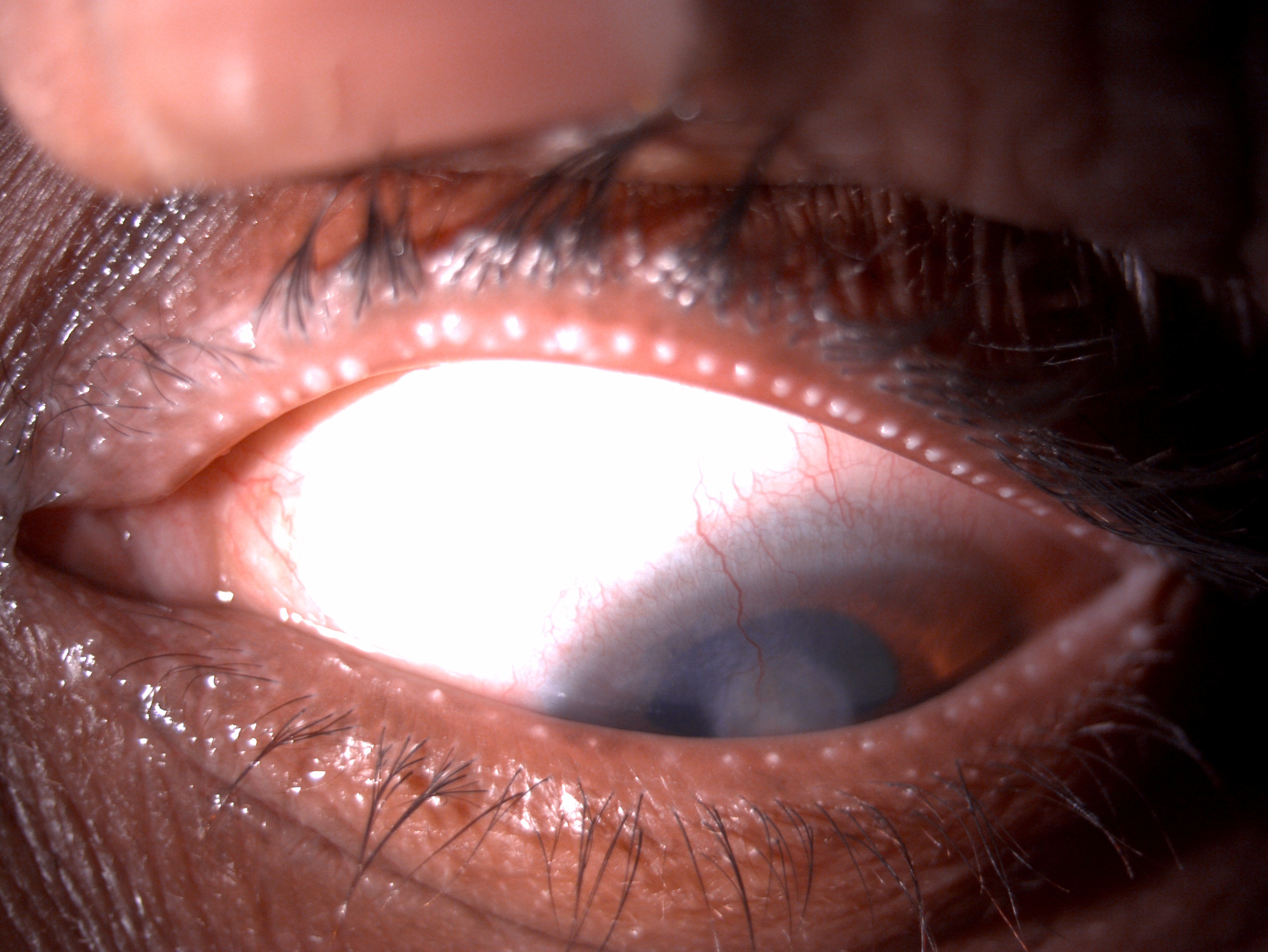
Digital slit lamp image of the patient depicting upper eyelid matted lashes, seborrheic blepharitis in lower eyelashes, upper
Digital slit lamp image of the patient depicting upper eyelid matted lashes, seborrheic blepharitis in lower eyelashes, upper lid margin blocked meibomian glands with sago grain appearance, corneal superficial vascularization along central nebular corneal opacity suggestive of MGD induced dry eye sequalae in the patient
Contributed by Dr. Kirandeep Kaur, MBBS, DNB, FPOS, FICO, MRCS Ed, MNAMS

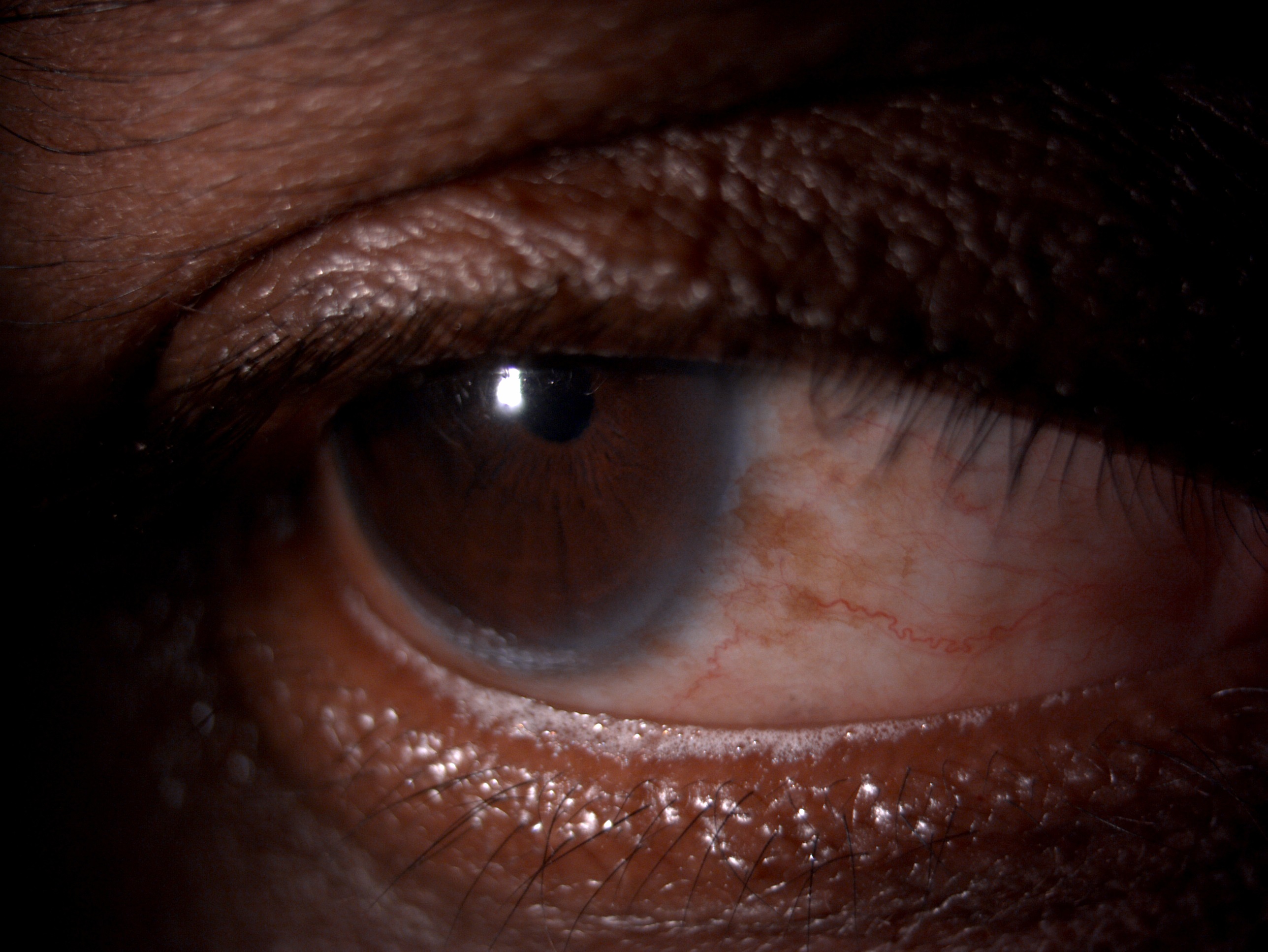
(Click Image to Enlarge)
Digital slit lamp image of the patient depicting frothing on the lower eyelid margin suggestive of meibomian gland disease
Contributed by Dr. Kirandeep Kaur, MBBS, DNB, FPOS, FICO, MRCS Ed, MNAMSd
Details

Editor:

Kirandeep Kaur

Updated:

12/6/2022 9:55:47 AM

References


[1]

Nelson JD, Shimazaki J, Benitez-del-Castillo JM, Craig JP, McCulley JP, Den S, Foulks GN. The international workshop on meibomian gland dysfunction: report of the definition and classification subcommittee. Investigative ophthalmology & visual science. 2011 Mar:52(4):1930-7. doi: 10.1167/iovs.10-6997b. Epub 2011 Mar 30     [PubMed PMID: 21450914]


[2]

Milner MS, Beckman KA, Luchs JI, Allen QB, Awdeh RM, Berdahl J, Boland TS, Buznego C, Gira JP, Goldberg DF, Goldman D, Goyal RK, Jackson MA, Katz J, Kim T, Majmudar PA, Malhotra RP, McDonald MB, Rajpal RK, Raviv T, Rowen S, Shamie N, Solomon JD, Stonecipher K, Tauber S, Trattler W, Walter KA, Waring GO 4th, Weinstock RJ, Wiley WF, Yeu E. Dysfunctional tear syndrome: dry eye disease and associated tear film disorders - new strategies for diagnosis and treatment. Current opinion in ophthalmology. 2017 Jan:27 Suppl 1(Suppl 1):3-47. doi: 10.1097/01.icu.0000512373.81749.b7. Epub     [PubMed PMID: 28099212]

Level 3 (low-level) evidence

[3]

Knop E, Knop N, Millar T, Obata H, Sullivan DA. The international workshop on meibomian gland dysfunction: report of the subcommittee on anatomy, physiology, and pathophysiology of the meibomian gland. Investigative ophthalmology & visual science. 2011 Mar:52(4):1938-78. doi: 10.1167/iovs.10-6997c. Epub 2011 Mar 30     [PubMed PMID: 21450915]


[4]

Tomlinson A, Bron AJ, Korb DR, Amano S, Paugh JR, Pearce EI, Yee R, Yokoi N, Arita R, Dogru M. The international workshop on meibomian gland dysfunction: report of the diagnosis subcommittee. Investigative ophthalmology & visual science. 2011 Mar:52(4):2006-49. doi: 10.1167/iovs.10-6997f. Epub 2011 Mar 30     [PubMed PMID: 21450918]


[5]

Chhadva P, Goldhardt R, Galor A. Meibomian Gland Disease: The Role of Gland Dysfunction in Dry Eye Disease. Ophthalmology. 2017 Nov:124(11S):S20-S26. doi: 10.1016/j.ophtha.2017.05.031. Epub     [PubMed PMID: 29055358]


[6]

Geerling G, Baudouin C, Aragona P, Rolando M, Boboridis KG, Benítez-Del-Castillo JM, Akova YA, Merayo-Lloves J, Labetoulle M, Steinhoff M, Messmer EM. Emerging strategies for the diagnosis and treatment of meibomian gland dysfunction: Proceedings of the OCEAN group meeting. The ocular surface. 2017 Apr:15(2):179-192. doi: 10.1016/j.jtos.2017.01.006. Epub 2017 Jan 27     [PubMed PMID: 28132878]


[7]

Putnam CM. Diagnosis and management of blepharitis: an optometrist's perspective. Clinical optometry. 2016:8():71-78. doi: 10.2147/OPTO.S84795. Epub 2016 Aug 8     [PubMed PMID: 30214351]

Level 3 (low-level) evidence

[8]

Schaumberg DA, Nichols JJ, Papas EB, Tong L, Uchino M, Nichols KK. The international workshop on meibomian gland dysfunction: report of the subcommittee on the epidemiology of, and associated risk factors for, MGD. Investigative ophthalmology & visual science. 2011 Mar:52(4):1994-2005. doi: 10.1167/iovs.10-6997e. Epub 2011 Mar 30     [PubMed PMID: 21450917]


[9]

Gurlevik U, Kemeriz F, Yasar E. The effect of isotretinoin on meibomian glands in eyes: a pilot study. International ophthalmology. 2022 Jul:42(7):2071-2078. doi: 10.1007/s10792-021-02205-1. Epub 2022 Jan 3     [PubMed PMID: 34978652]

Level 3 (low-level) evidence

[10]

Hassanzadeh S, Varmaghani M, Zarei-Ghanavati S, Heravian Shandiz J, Azimi Khorasani A. Global Prevalence of Meibomian Gland Dysfunction: A Systematic Review and Meta-Analysis. Ocular immunology and inflammation. 2021 Jan 2:29(1):66-75. doi: 10.1080/09273948.2020.1755441. Epub 2020 Jun 26     [PubMed PMID: 32589483]

Level 1 (high-level) evidence

[11]

Alghamdi YA, Mercado C, McClellan AL, Batawi H, Karp CL, Galor A. Epidemiology of Meibomian Gland Dysfunction in an Elderly Population. Cornea. 2016 Jun:35(6):731-5. doi: 10.1097/ICO.0000000000000815. Epub     [PubMed PMID: 27027919]


[12]

Badian RA, Utheim TP, Chen X, Utheim ØA, Ræder S, Ystenæs AE, Aakre BM, Sundling V. Meibomian gland dysfunction is highly prevalent among first-time visitors at a Norwegian dry eye specialist clinic. Scientific reports. 2021 Dec 3:11(1):23412. doi: 10.1038/s41598-021-02738-6. Epub 2021 Dec 3     [PubMed PMID: 34862425]


[13]

Suzuki T. Inflamed Obstructive Meibomian Gland Dysfunction Causes Ocular Surface Inflammation. Investigative ophthalmology & visual science. 2018 Nov 1:59(14):DES94-DES101. doi: 10.1167/iovs.17-23345. Epub     [PubMed PMID: 30481812]


[14]

Messmer EM. The pathophysiology, diagnosis, and treatment of dry eye disease. Deutsches Arzteblatt international. 2015 Jan 30:112(5):71-81; quiz 82. doi: 10.3238/arztebl.2015.0071. Epub     [PubMed PMID: 25686388]


[15]

Gutgesell VJ,Stern GA,Hood CI, Histopathology of meibomian gland dysfunction. American journal of ophthalmology. 1982 Sep;     [PubMed PMID: 7124880]


[16]

Obata H. Anatomy and histopathology of human meibomian gland. Cornea. 2002 Oct:21(7 Suppl):S70-4     [PubMed PMID: 12484702]


[17]

Ha M, Kim JS, Hong SY, Chang DJ, Whang WJ, Na KS, Kim EC, Kim HS, Hwang HS. Relationship between eyelid margin irregularity and meibomian gland dropout. The ocular surface. 2021 Jan:19():31-37. doi: 10.1016/j.jtos.2020.11.007. Epub 2020 Nov 24     [PubMed PMID: 33246034]


[18]

Sabeti S, Kheirkhah A, Yin J, Dana R. Management of meibomian gland dysfunction: a review. Survey of ophthalmology. 2020 Mar-Apr:65(2):205-217. doi: 10.1016/j.survophthal.2019.08.007. Epub 2019 Sep 5     [PubMed PMID: 31494111]

Level 3 (low-level) evidence

[19]

Yokoi N,Bron AJ,Tiffany JM,Maruyama K,Komuro A,Kinoshita S, Relationship between tear volume and tear meniscus curvature. Archives of ophthalmology (Chicago, Ill. : 1960). 2004 Sep;     [PubMed PMID: 15364704]


[20]

Robin M, Liang H, Rabut G, Augstburger E, Baudouin C, Labbé A. The Role of Meibography in the Diagnosis of Meibomian Gland Dysfunction in Ocular Surface Diseases. Translational vision science & technology. 2019 Nov:8(6):6. doi: 10.1167/tvst.8.6.6. Epub 2019 Nov 12     [PubMed PMID: 31737430]


[21]

Wise RJ, Sobel RK, Allen RC. Meibography: A review of techniques and technologies. Saudi journal of ophthalmology : official journal of the Saudi Ophthalmological Society. 2012 Oct:26(4):349-56. doi: 10.1016/j.sjopt.2012.08.007. Epub     [PubMed PMID: 23961019]


[22]

Lee JM,Jeon YJ,Kim KY,Hwang KY,Kwon YA,Koh K, Ocular surface analysis: A comparison between the LipiView{sup}®{/sup} II and IDRA{sup}®{/sup}. European journal of ophthalmology. 2021 Sep;     [PubMed PMID: 33267698]


[23]

Arita R, Fukuoka S, Kawashima M. Proposed Algorithm for Management of Meibomian Gland Dysfunction Based on Noninvasive Meibography. Journal of clinical medicine. 2020 Dec 27:10(1):. doi: 10.3390/jcm10010065. Epub 2020 Dec 27     [PubMed PMID: 33375436]


[24]

Xiao J, Adil MY, Chen X, Utheim ØA, Ræder S, Tønseth KA, Lagali NS, Dartt DA, Utheim TP. Functional and Morphological Evaluation of Meibomian Glands in the Assessment of Meibomian Gland Dysfunction Subtype and Severity. American journal of ophthalmology. 2020 Jan:209():160-167. doi: 10.1016/j.ajo.2019.09.005. Epub 2019 Sep 14     [PubMed PMID: 31526799]


[25]

Paugh JR, Kwan J, Christensen M, Nguyen AL, Senchyna M, Meadows D. Development of a Meibomian Gland Dysfunction-Specific Symptom Questionnaire. Eye & contact lens. 2018 Jan:44(1):6-14. doi: 10.1097/ICL.0000000000000294. Epub     [PubMed PMID: 27466720]


[26]

Okumura Y, Inomata T, Iwata N, Sung J, Fujimoto K, Fujio K, Midorikawa-Inomata A, Miura M, Akasaki Y, Murakami A. A Review of Dry Eye Questionnaires: Measuring Patient-Reported Outcomes and Health-Related Quality of Life. Diagnostics (Basel, Switzerland). 2020 Aug 5:10(8):. doi: 10.3390/diagnostics10080559. Epub 2020 Aug 5     [PubMed PMID: 32764273]

Level 2 (mid-level) evidence

[27]

Nichols KK,Foulks GN,Bron AJ,Glasgow BJ,Dogru M,Tsubota K,Lemp MA,Sullivan DA, The international workshop on meibomian gland dysfunction: executive summary. Investigative ophthalmology     [PubMed PMID: 21450913]


[28]

McCulley JP, Shine WE. The lipid layer of tears: dependent on meibomian gland function. Experimental eye research. 2004 Mar:78(3):361-5     [PubMed PMID: 15106913]


[29]

Kobayashi A, Ide T, Fukumoto T, Miki E, Tsubota K, Toda I. Effects of a New Eyelid Shampoo on Lid Hygiene and Eyelash Length in Patients with Meibomian Gland Dysfunction: A Comparative Open Study. Journal of ophthalmology. 2016:2016():4292570. doi: 10.1155/2016/4292570. Epub 2016 Dec 19     [PubMed PMID: 28078136]

Level 2 (mid-level) evidence

[30]

Kocamiş Ö, Temel E, Aşikgarip N, Örnek K. Electronic Device Screen Time and Meibomian Gland Morphology in Children. Journal of ophthalmic & vision research. 2021 Oct-Dec:16(4):531-537. doi: 10.18502/jovr.v16i4.9741. Epub 2021 Oct 25     [PubMed PMID: 34840674]


[31]

Macsai MS, The role of omega-3 dietary supplementation in blepharitis and meibomian gland dysfunction (an AOS thesis). Transactions of the American Ophthalmological Society. 2008;     [PubMed PMID: 19277245]


[32]

Oleñik A, Mahillo-Fernández I, Alejandre-Alba N, Fernández-Sanz G, Pérez MA, Luxan S, Quintana S, Martínez de Carneros Llorente A, García-Sandoval B, Jiménez-Alfaro I. Benefits of omega-3 fatty acid dietary supplementation on health-related quality of life in patients with meibomian gland dysfunction. Clinical ophthalmology (Auckland, N.Z.). 2014:8():831-6. doi: 10.2147/OPTH.S62470. Epub 2014 Apr 30     [PubMed PMID: 24812490]

Level 2 (mid-level) evidence

[33]

Garrigue JS, Amrane M, Faure MO, Holopainen JM, Tong L. Relevance of Lipid-Based Products in the Management of Dry Eye Disease. Journal of ocular pharmacology and therapeutics : the official journal of the Association for Ocular Pharmacology and Therapeutics. 2017 Nov:33(9):647-661. doi: 10.1089/jop.2017.0052. Epub 2017 Sep 28     [PubMed PMID: 28956698]


[34]

Kim HY, Lee JE, Oh HN, Song JW, Han SY, Lee JS. Clinical efficacy of combined topical 0.05% cyclosporine A and 0.1% sodium hyaluronate in the dry eyes with meibomian gland dysfunction. International journal of ophthalmology. 2018:11(4):593-600. doi: 10.18240/ijo.2018.04.09. Epub 2018 Apr 18     [PubMed PMID: 29675376]


[35]

Qiao J, Yan X. Emerging treatment options for meibomian gland dysfunction. Clinical ophthalmology (Auckland, N.Z.). 2013:7():1797-803. doi: 10.2147/OPTH.S33182. Epub 2013 Sep 9     [PubMed PMID: 24043929]

Level 2 (mid-level) evidence

[36]

Maskin SL. Intraductal meibomian gland probing relieves symptoms of obstructive meibomian gland dysfunction. Cornea. 2010 Oct:29(10):1145-52. doi: 10.1097/ICO.0b013e3181d836f3. Epub     [PubMed PMID: 20622668]


[37]

Magno M,Moschowits E,Arita R,Vehof J,Utheim TP, Intraductal meibomian gland probing and its efficacy in the treatment of meibomian gland dysfunction. Survey of ophthalmology. 2021 Jul-Aug;     [PubMed PMID: 33352147]

Level 3 (low-level) evidence

[38]

Syed ZA, Sutula FC. Dynamic Intraductal Meibomian Probing: A Modified Approach to the Treatment of Obstructive Meibomian Gland Dysfunction. Ophthalmic plastic and reconstructive surgery. 2017 Jul/Aug:33(4):307-309. doi: 10.1097/IOP.0000000000000876. Epub     [PubMed PMID: 28221295]


[39]

Lane SS, DuBiner HB, Epstein RJ, Ernest PH, Greiner JV, Hardten DR, Holland EJ, Lemp MA, McDonald JE 2nd, Silbert DI, Blackie CA, Stevens CA, Bedi R. A new system, the LipiFlow, for the treatment of meibomian gland dysfunction. Cornea. 2012 Apr:31(4):396-404. doi: 10.1097/ICO.0b013e318239aaea. Epub     [PubMed PMID: 22222996]


[40]

Greiner JV. Long-Term (3 Year) Effects of a Single Thermal Pulsation System Treatment on Meibomian Gland Function and Dry Eye Symptoms. Eye & contact lens. 2016 Mar:42(2):99-107. doi: 10.1097/ICL.0000000000000166. Epub     [PubMed PMID: 26222095]


[41]

Finis D, König C, Hayajneh J, Borrelli M, Schrader S, Geerling G. Six-month effects of a thermodynamic treatment for MGD and implications of meibomian gland atrophy. Cornea. 2014 Dec:33(12):1265-70. doi: 10.1097/ICO.0000000000000273. Epub     [PubMed PMID: 25321941]


[42]

Li S, Yang K, Wang J, Li S, Zhu L, Feng J, Tian L, Jie Y. Effect of a Novel Thermostatic Device on Meibomian Gland Dysfunction: A Randomized Controlled Trial in Chinese Patients. Ophthalmology and therapy. 2022 Feb:11(1):261-270. doi: 10.1007/s40123-021-00431-5. Epub 2021 Nov 25     [PubMed PMID: 34822140]

Level 1 (high-level) evidence

[43]

Goldberg DJ. Current trends in intense pulsed light. The Journal of clinical and aesthetic dermatology. 2012 Jun:5(6):45-53     [PubMed PMID: 22768357]


[44]

Bäumler W, Vural E, Landthaler M, Muzzi F, Shafirstein G. The effects of intense pulsed light (IPL) on blood vessels investigated by mathematical modeling. Lasers in surgery and medicine. 2007 Feb:39(2):132-9     [PubMed PMID: 17066482]


[45]

Brinton M, Kossler AL, Patel ZM, Loudin J, Franke M, Ta CN, Palanker D. Enhanced Tearing by Electrical Stimulation of the Anterior Ethmoid Nerve. Investigative ophthalmology & visual science. 2017 Apr 1:58(4):2341-2348. doi: 10.1167/iovs.16-21362. Epub     [PubMed PMID: 28431436]


[46]

Bhandari V, Reddy JK. Blepharitis: always remember demodex. Middle East African journal of ophthalmology. 2014 Oct-Dec:21(4):317-20. doi: 10.4103/0974-9233.142268. Epub     [PubMed PMID: 25371637]


[47]

Kassan SS,Moutsopoulos HM, Clinical manifestations and early diagnosis of Sjögren syndrome. Archives of internal medicine. 2004 Jun 28;     [PubMed PMID: 15226160]


[48]

Vazirani J, Donthineni PR, Goel S, Sane SS, Mahuvakar S, Narang P, Shanbhag SS, Basu S. Chronic cicatrizing conjunctivitis: A review of the differential diagnosis and an algorithmic approach to management. Indian journal of ophthalmology. 2020 Nov:68(11):2349-2355. doi: 10.4103/ijo.IJO_604_20. Epub     [PubMed PMID: 33120615]


[49]

Zhang XB, Ding YH, He W. The association between demodex infestation and ocular surface manifestations in meibomian gland dysfunction. International journal of ophthalmology. 2018:11(4):589-592. doi: 10.18240/ijo.2018.04.08. Epub 2018 Apr 18     [PubMed PMID: 29675375]


[50]

Jiang X, Deng A, Yang J, Bai H, Yang Z, Wu J, Lv H, Li X, Wen T. Pathogens in the Meibomian gland and conjunctival sac: microbiome of normal subjects and patients with Meibomian gland dysfunction. Infection and drug resistance. 2018:11():1729-1740. doi: 10.2147/IDR.S162135. Epub 2018 Oct 11     [PubMed PMID: 30349330]


[51]

Arita R, Fukuoka S. Non-pharmaceutical treatment options for meibomian gland dysfunction. Clinical & experimental optometry. 2020 Nov:103(6):742-755. doi: 10.1111/cxo.13035. Epub 2020 Jan 13     [PubMed PMID: 31943385]


[52]

Yin Y,Gong L, Reversibility of Gland Dropout and Significance of Eyelid Hygiene Treatment in Meibomian Gland Dysfunction. Cornea. 2017 Mar;     [PubMed PMID: 27755193]


[53]

Downie LE, Craig JP. Tear film evaluation and management in soft contact lens wear: a systematic approach. Clinical & experimental optometry. 2017 Sep:100(5):438-458. doi: 10.1111/cxo.12597. Epub     [PubMed PMID: 28940531]

Level 1 (high-level) evidence

[54]

Mizoguchi S, Iwanishi H, Arita R, Shirai K, Sumioka T, Kokado M, Jester JV, Saika S. Ocular surface inflammation impairs structure and function of meibomian gland. Experimental eye research. 2017 Oct:163():78-84. doi: 10.1016/j.exer.2017.06.011. Epub     [PubMed PMID: 28950941]


[55]

Hau S, Ehrlich D, Binstead K, Verma S. An evaluation of optometrists' ability to correctly identify and manage patients with ocular disease in the accident and emergency department of an eye hospital. The British journal of ophthalmology. 2007 Apr:91(4):437-40     [PubMed PMID: 17077113]