Introduction
Chronic cholecystitis is a chronic condition caused by ongoing inflammation of the gallbladder resulting in mechanical or physiological dysfunction its emptying. It presents as a smoldering course that can be accompanied by acute exacerbations of increased pain (acute biliary colic), or it can progress to a more severe form of cholecystitis requiring urgent intervention (acute cholecystitis). There are classic signs and symptoms associated with this disease as well as prevalence in certain patient populations. The two forms of chronic cholecystitis are calculous (occuring in the setting of cholelithiasis), and acalculous (without gallstones). However most cases of chronic cholecystitis are commonly associated with cholelithiasis.
Etiology
Register For Free And Read The Full Article
Search engine and full access to all medical articles
10 free questions in your specialty
Free CME/CE Activities
Free daily question in your email
Save favorite articles to your dashboard
Emails offering discounts
Learn more about a Subscription to StatPearls Point-of-Care
Etiology
Chronic cholecystitis mostly occurs in the setting of cholelithiasis. The proposed etiology is recurrent episodes of acute cholecystitis or chronic irritation from gallstones invoking an inflammatory response in the gallbladder wall. Sometimes the term is used to describe abdominal pain resulting from dysfunction in the emptying of the gallbladder. This overlaps with Sphincter of Oddi dysfunction and is best referred to as biliary or gallbladder dyskinesia.
Risk factors for cholelithiasis include:
- Female gender
- Obesity
- Rapid weight loss
- Pregnancy
- Advanced age
- Hispanic or Pima Indians
Epidemiology
The epidemiology of chronic cholecystitis mostly parallels with that of cholelithiasis. Specific data on this disease entity is limited.
Gallstone disease is very common. About 10-20% of the world population will develop gallstones at some point in their life and about 80% of them are asymptomatic[1]. There are approximately 500,000 cholecystectomies done yearly in the United Stated for gallbladder disease. The incidence of gallstone formation increases yearly with age. Over one-quarter of women older than the age of 60 will have gallstones. In the United States, approximately 14 million women and 6 million men with an age range of 20 to 74 have gallstones. Obesity increases the likelihood of gallstones, especially in women due to increases in the biliary secretion of cholesterol. On the other hand, patients with drastic weight loss or fasting have a higher chance of gallstones secondary to biliary stasis. Furthermore, there is also a hormonal association with gallstones. Estrogen has been shown to result in an increase in bile cholesterol as well as a decrease in gallbladder contractility. Women of reproductive age or on estrogen-containing contraceptives have a two-fold increase in gallstone formation compared to males. People with chronic illnesses such as diabetes also have an increase in gallstone formation as well as reduced gallbladder wall contractility due to neuropathy.[2]
Pathophysiology
Occlusion of the cystic duct or malfunction of the mechanics of the gallbladder emptying is the basic underlying pathologies of this disease. Over 90% of chronic cholecystitis is associated with the presence of gallstones. Gallstones, by causing intermittent obstruction of the bile flow, most commonly by blocking the cystic duct lead to inflammation and edema in the gall bladder wall. Occlusion of the common bile duct such as in neoplasms or strictures can also lead to stasis of the bile flow causing gallstone formation with resultant chronic cholecystitis.[3]
It has been proposed that lithogenic bile leads to increased free radical-mediated damage from hydrophobic bile salts. That, in association with reduced mucosal protection due to lower levels of prostaglandin E2 results in a continuous inflammatory state. When the cholecystokinin receptors of the smooth muscle are affected, there is impaired gall bladder contraction that leads to stasis and worsens the permissive environment where lithogenic bile promotes inflammation.[4]
Histopathology
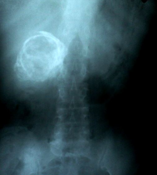
The gallbladder wall may be thickened to variable degrees, and there may be adhesions to the serosal surface. In some cases, due to extensive fibrosis, the gallbladder may appear shrunken. Smooth muscle hypertrophy, especially in prolonged chronic conditions, is present. Calcium bilirubinate or cholesterol stones are most often present and can vary in size from sand-like to completely filling the entire gallbladder lumen. They can be multiple or singular. The acalculous disease may reveal sludge or very viscous bile. These findings are usual precursors to gallstones and are formed from increased biliary salts or stasis. Normal appearing bile can also be present. Various species of bacteria can be found in 11% to 30% of the cases. Rokitansky-Aschoff sinuses are present or accentuated in 90% of the time in chronic cholecystitis specimens. These are a herniation of intraluminal sinuses from increased pressures possibly associated with ducts of Luschka. The mucosa will exhibit varying degrees of inflammation. T lymphocytes are the common cells followed by plasma cells and histiocytes. Metaplastic changes can be seen. There is usually hypertrophy of the muscularis mucosa with varying degrees of mural fibrosis and elastosis. A variant in which calcium deposition and hyaline fibrosis leads to diffuse thinning of the gallbladder wall is called hyalinizing cholecystitis. The brittle consistency also gives it the name porcelain gallbladder.[5]
History and Physical
Symptomatic patients with chronic cholecystitis usually present with dull right upper abdominal pain that radiates around the waist to the mid back or right scapular tip. The pain may be exacerbated by fatty food intake but the classical post-prandial pain of acute cholecystitis is less common. Nausea and occasional vomiting also accompany complaints of increased bloating and flatulence. Often the symptoms occur in the evening or at night. Symptoms are usually present over weeks to months as opposed to the abrupt, severe presentation of acute cholecystitis. There might be a gradual worsening of symptoms or an increase in the frequency of episodes. Fever and tachycardia are rare. Elderly patients with cholecystitis may present with vague symptoms and they are at risk of progression to complicated disease. Hence a high index of clinical suspicion is required in the diagnosis of this condition.
Evaluation
Laboratory testing is not specific or sensitive in making a diagnosis of chronic cholecystitis. Leukocytosis and abnormal liver function tests may not be present in these patients, unlike the acute disease. However basic laboratory testing in the form of a metabolic panel, liver functions, and complete blood count should be performed. Cardiac testing including EKG and troponins should be considered in the appropriate clinical setting.
The diagnostic investigation of choice when chronic cholecystitis is suspected clinically is a right upper quadrant ultrasound. This non-invasive study that is readily available in most facilities can accurately evaluate the gallbladder for a thickened wall or inflammation. It also aids in the evaluation of gallstones or sludge. Computerized tomography (CT) with intravenous contrast usually reveals cholelithiasis, increased attenuation of bile, and gallbladder wall thickening. The gallbladder itself may appear distended or contracted, however, pericholecystic inflammation and fluid collection are usually absent.[6] A distended gallbladder and increased enhancement of adjacent hepatic tissue go more in favor of acute cholecystitis, whereas hyperenhancement of the gallbladder wall is more commonly seen in the chronic disease.[7] Given the overlapping findings between acute and chronic cholecystitis, sometimes ultrasound and CT may be adequate to come to a final diagnosis. A magnetic resonance imaging (MRI) study is a useful alternative in patients who are unable to undergo a CT scan due to radiation concerns or renal injury.[8] The diagnostic test of choice to confirm chronic cholecystitis is the hepatobiliary scintigraphy or a HIDA scan with cholecystokinin(CCK). The most common scintigraphic findings are delayed gallbladder visualization (between 1-4 hours) and delayed increased biliary to bowel transit time.[9] The tracer is injected intravascularly and gets concentrated in the gallbladder. CCK is then administered and the percentage of gallbladder emptying (ejection fraction - EF) is calculated. An EF below 35% at the 15-minute cutoff is considered a dyskinetic gallbladder and is suggestive of chronic cholecystitis.
Treatment / Management
The preferred treatment for chronic cholecystitis is elective laparoscopic cholecystectomy. It has a low morbidity rate and can be performed as an outpatient surgery. An open cholecystectomy is also an option however requires hospital admission and longer recovery time. This surgery is indicated in patients who are not laparoscopic candidates such as those with extensive prior surgeries and adhesions. Endoscopic retrograde cholangiopancreatography (ERCP) is usually done when choledocholithiasis is a concern. These patients usually undergo ERCP prior to elective surgery.
Patients who are not surgical candidates or who prefer not to undergo surgery can be closely observed and managed conservatively. A low-fat diet can help reduce the frequency of symptoms. In patients with symptomatic cholelithiasis, the use of ursodeoxycholic acid (UDCA or ursodiol) has been shown to decrease rates of biliary colic and acute cholecystitis.[10] However, the literature on its role in chronic cholecystitis is limited. The management of asymptomatic patients with incidentally detected chronic cholecystitis depends on patient characteristics. Asymptomatic patients with no radiological or clinical concerns of malignancy can also be closely monitored with follow-up imaging.
Differential Diagnosis
There are other common medical conditions that can mimic the presentation of chronic cholecystitis. A thorough analysis of the clinical presentation often can guide appropriate workup. Common clinical features of these disorders are as follows:
- Acute cholecystitis: A continuous, severe pain in the right side of the abdomen lasting for hours associated with fever, nausea, and vomiting in an ill-looking patient is suggestive of acute cholecystitis[11].
- Gall bladder cancer: Chronic abdominal symptoms associated with weight loss or other constitutional symptoms should raise suspicion of this. Imaging and histology are helpful in making a definitive diagnosis.
- Peptic ulcer disease: The presence of epigastric abdominal pain and early satiety should alert the possibility of peptic ulcer disease. Alarm symptoms include weight loss, anemia, melena or dysphagia[12].
- GERD: Burning sensation in the epigastrium or retrosternal region that may be associated with regurgitation of food material.
- Gastric cancer: the presence of alarm symptoms of peptic ulcer disease, persistent vomiting, evidence of malignancy or other risk factors should alert to the possibility of this[13].
- Myocardial infarction: In cases of the inferior wall or right ventricular ischemia, the presenting symptoms may be epigastric pain with nausea and vomiting. Other cardiac symptoms like dizziness or SOB or risk factors for coronary ischemia should prompt a workup for the same[14].
- Mesenteric ischemia: the acute variant presents with severe acute abdominal pain and the chronic variant typically with post-prandial pain. Old age, risk factors for atherosclerosis, blood in stools, and weight loss are concerning features of this condition[15].
- Mesenteric vasculitis: presence of ongoing abdominal symptoms unexplained by regular workup and the presence of other features consistent with systemic vasculitis could be related to this relatively underrecognized but dangerous condition[16].
Prognosis
The majority of uncomplicated cases of cholecystitis have an excellent outcome. In many cases, supportive treatments can help with symptoms. Most cases are treated with elective cholecystectomy to prevent future complications. While surgery is safe, bile duct injuries can happen and need to be monitored in the post-operative period.
Complications
The proliferation of bacteria in the gallbladder can lead to acute cholecystitis or pus collections. In some cases, the gallstone may erode into the duodenum and impact in the terminal ileum, presenting as gallstone ileus. Rarely the patient may develop emphysematous cholecystitis due to the presence of gas-forming organisms like clostridia, E.coli, and klebsiella. This presentation is most common in diabetics and carries a high mortality rate. The relationship between chronic cholecystitis and gall bladder cancer is controversial. Though chronic inflammation has been shown to be associated with increased risk of cancer[17], the data on this is limited. Xanthogranulomatous cholecystitis is a variant of chronic cholecystitis in which continued inflammation leads to extensive thickening and fibrosis extending locally beyond the gall bladder wall. In this severe variant, the occurrence of complications like abscesses and fistulas are more common. It is considered a pre-malignant condition. Porcelain gallbladder tends to be asymptomatic in most cases. The association with malignancy is again controversial but the consensus is that it carries a slightly increased risk of cancer.[18]
Consultations
The diagnosis is usually made at the level of primary care or in the inpatient setting. A gastroenterology consult is mandated when gallstone obstruction of the biliary system is suspected. Otherwise, most patients are referred to general surgery for consideration of elective cholecystectomy.
Enhancing Healthcare Team Outcomes
The diagnosis and management of cholecystitis is a multi-disciplinary team approach. A high index of suspicion is vital in the diagnosis. Referral to the surgical team followed by decision making on the need for laparoscopic surgery are the next steps. Good surgical care with good postoperative follow up is also essential. Counseling for food habits with nutritionist support and lifestyle changes are crucial in patients being treated conservatively.
Media
References
Stinton LM, Shaffer EA. Epidemiology of gallbladder disease: cholelithiasis and cancer. Gut and liver. 2012 Apr:6(2):172-87. doi: 10.5009/gnl.2012.6.2.172. Epub 2012 Apr 17 [PubMed PMID: 22570746]
Andercou O, Olteanu G, Mihaileanu F, Stancu B, Dorin M. Risk factors for acute cholecystitis and for intraoperative complications. Annali italiani di chirurgia. 2017:88():318-325 [PubMed PMID: 29068324]
Wang L, Sun W, Chang Y, Yi Z. Differential proteomics analysis of bile between gangrenous cholecystitis and chronic cholecystitis. Medical hypotheses. 2018 Dec:121():131-136. doi: 10.1016/j.mehy.2018.07.004. Epub 2018 Jul 5 [PubMed PMID: 30396466]
Guarino MP, Cong P, Cicala M, Alloni R, Carotti S, Behar J. Ursodeoxycholic acid improves muscle contractility and inflammation in symptomatic gallbladders with cholesterol gallstones. Gut. 2007 Jun:56(6):815-20 [PubMed PMID: 17185355]
Level 1 (high-level) evidenceBenkhadoura M, Elshaikhy A, Eldruki S, Elfaedy O. Routine histopathological examination of gallbladder specimens after cholecystectomy: Is it time to change the current practice? Turkish journal of surgery. 2018 Sep 11:():1-4. doi: 10.5152/turkjsurg.2018.4126. Epub 2018 Sep 11 [PubMed PMID: 30248293]
Smith EA, Dillman JR, Elsayes KM, Menias CO, Bude RO. Cross-sectional imaging of acute and chronic gallbladder inflammatory disease. AJR. American journal of roentgenology. 2009 Jan:192(1):188-96. doi: 10.2214/AJR.07.3803. Epub [PubMed PMID: 19098200]
Level 2 (mid-level) evidenceYeo DM, Jung SE. Differentiation of acute cholecystitis from chronic cholecystitis: Determination of useful multidetector computed tomography findings. Medicine. 2018 Aug:97(33):e11851. doi: 10.1097/MD.0000000000011851. Epub [PubMed PMID: 30113479]
Kaura SH,Haghighi M,Matza BW,Hajdu CH,Rosenkrantz AB, Comparison of CT and MRI findings in the differentiation of acute from chronic cholecystitis. Clinical imaging. 2013 Jul-Aug [PubMed PMID: 23541278]
Level 3 (low-level) evidenceChamarthy M, Freeman LM. Hepatobiliary scan findings in chronic cholecystitis. Clinical nuclear medicine. 2010 Apr:35(4):244-51. doi: 10.1097/RLU.0b013e3181d18ef5. Epub [PubMed PMID: 20305411]
Guarino MP, Cocca S, Altomare A, Emerenziani S, Cicala M. Ursodeoxycholic acid therapy in gallbladder disease, a story not yet completed. World journal of gastroenterology. 2013 Aug 21:19(31):5029-34. doi: 10.3748/wjg.v19.i31.5029. Epub [PubMed PMID: 23964136]
Huffman JL, Schenker S. Acute acalculous cholecystitis: a review. Clinical gastroenterology and hepatology : the official clinical practice journal of the American Gastroenterological Association. 2010 Jan:8(1):15-22. doi: 10.1016/j.cgh.2009.08.034. Epub 2009 Sep 10 [PubMed PMID: 19747982]
Malik TF,Gnanapandithan K,Singh K, Peptic Ulcer Disease . 2020 Jan [PubMed PMID: 30521213]
Ajani JA, Lee J, Sano T, Janjigian YY, Fan D, Song S. Gastric adenocarcinoma. Nature reviews. Disease primers. 2017 Jun 1:3():17036. doi: 10.1038/nrdp.2017.36. Epub 2017 Jun 1 [PubMed PMID: 28569272]
Albulushi A, Giannopoulos A, Kafkas N, Dragasis S, Pavlides G, Chatzizisis YS. Acute right ventricular myocardial infarction. Expert review of cardiovascular therapy. 2018 Jul:16(7):455-464. doi: 10.1080/14779072.2018.1489234. Epub 2018 Jun 27 [PubMed PMID: 29902098]
Gnanapandithan K, Feuerstadt P. Review Article: Mesenteric Ischemia. Current gastroenterology reports. 2020 Mar 17:22(4):17. doi: 10.1007/s11894-020-0754-x. Epub 2020 Mar 17 [PubMed PMID: 32185509]
Gnanapandithan K,Sharma A, Mesenteric Vasculitis . 2020 Jan [PubMed PMID: 31536217]
Goetze TO. Gallbladder carcinoma: Prognostic factors and therapeutic options. World journal of gastroenterology. 2015 Nov 21:21(43):12211-7. doi: 10.3748/wjg.v21.i43.12211. Epub [PubMed PMID: 26604631]
Patel S, Roa JC, Tapia O, Dursun N, Bagci P, Basturk O, Cakir A, Losada H, Sarmiento J, Adsay V. Hyalinizing cholecystitis and associated carcinomas: clinicopathologic analysis of a distinctive variant of cholecystitis with porcelain-like features and accompanying diagnostically challenging carcinomas. The American journal of surgical pathology. 2011 Aug:35(8):1104-13. doi: 10.1097/PAS.0b013e31822179cc. Epub [PubMed PMID: 21716080]
