Introduction
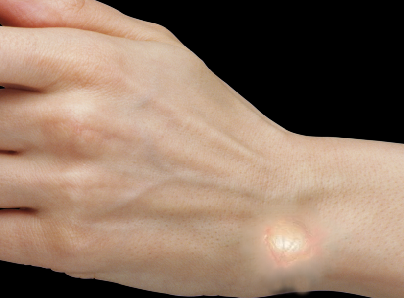
Ganglion cysts are synovial cysts that are filled with gelatinous mucoid material and commonly encountered in orthopedic clinical practice. Although the exact etiology of the development of ganglion cysts is unknown, they are believed to arise from repetitive microtrauma resulting in mucinous degeneration of connective tissue. They are the most common soft tissue mass found within the hand and wrist, but they are also commonly encountered in the knee and foot. Although the majority of ganglion cysts are asymptomatic, patients may present with pain, tenderness, weakness, and dissatisfaction with cosmetic appearance. Both non-operative and surgical treatments are available, but a high recurrence rate has historically plagued non-surgical treatment. Surgical excision can provide resolution of patients' symptoms, but an understanding of the underlying anatomy adjacent to the cyst is crucial to avoid injuring neurovascular structures within proximity to the cyst.[1][2]
Etiology
Register For Free And Read The Full Article
Search engine and full access to all medical articles
10 free questions in your specialty
Free CME/CE Activities
Free daily question in your email
Save favorite articles to your dashboard
Emails offering discounts
Learn more about a Subscription to StatPearls Point-of-Care
Etiology
Numerous theories have been presented in the past regarding the etiology of ganglion cysts with no present consensus. One theory introduced by Eller in 1746 is that ganglion cysts are the result of the herniation of synovial tissue from joints. Another theory postulated by Carp and Stout in 1926, which forms the basis of most modern belief, suggests that ganglion cysts result from mucinous degeneration of connective tissue secondary to chronic damage. Currently, most authors agree that ganglion cysts arise from mesenchymal cells at the synovial capsular junction as a result of the continuous micro-injury. Repetitive injury to the supporting capsular and ligamentous structures appears to stimulate fibroblasts to produce hyaluronic acid, which accumulates to produce the mucin "jelly-like" material commonly found in ganglion cysts.[3][4][5]
Epidemiology
Ganglion cysts account for 60% to 70% of soft-tissue masses found in the hand and wrist. Although they can form at any age, they are most commonly found in women between the ages of 20 to 50. Women are three times more likely to develop a ganglion cyst than men. These cysts are also frequently encountered amongst gymnasts, likely secondary to repetitive trauma and stress of the wrist joint.
Pathophysiology
Ganglion cysts are mucin filled synovial cysts containing paucicellular connective tissue. They may be filled with fluid from a tendon sheath or joint. Ganglion cysts are most commonly found (70%) on the dorsal aspect of the wrist arising from the scapholunate ligament or scapholunate articulation. Approximately 20% of ganglion cysts are located on the volar aspect of the wrist arising from the radiocarpal joint or scaphotrapezial joint. The remaining 10% of ganglion cysts can arise from multiple areas of the body including the volar retinaculum of the wrist, distal interphalangeal joint, ankle joint, and foot. Wrist volar retinacular cysts arise from herniated tendon sheath fluid that protrudes out. Ganglion cysts arising from the dorsal DIP joint are called mucous cysts and are associated with Herbeden's nodules. They are commonly found in women between the ages of 40 and 70 with osteoarthritis.[6][7][8]
Histopathology
Biopsies of ganglion cysts are not routinely indicated because of their inherently benign nature. Typical histopathological appearance is a mucin-filled synovial cell lined sac without a true epithelial lining. Ganglion cysts can be single or multiloculated. When examined under electron microscopy, their walls contain sheets of collagen fibers arranged in multidirectional layers with intermittent flattened cells resembling fibroblasts. The thick mucinous material present in the majority of ganglion cysts is highly viscous, which is attributed to a high concentration of hyaluronic acid and mucopolysaccharides.
History and Physical
The majority of ganglion cysts are asymptomatic, but patients may seek treatment because of their unsightly cosmetic appearance. Patients may present with pain, tenderness, or weakness that is exacerbated by wrist motion. Ganglion cysts usually present as firm, well circumscribed, freely mobile masses approximately 1 cm to 3 cm in size. They are often fixed to deep tissue and not to the overlying skin. Patients with volar wrist ganglion cysts less commonly may present with carpal tunnel syndrome or a trigger finger secondary to compression of the median nerve or intrusion on the flexor tendon sheath. Volar wrist ganglion cysts can also cause ulnar nerve neuropraxia and compression of the radial artery leading to ischemia. Ganglion cysts will usually transilluminate on the exam.
Evaluation
Radiographs may be ordered to rule out any related intraosseous manifestation, but will generally be unremarkable. MRI is usually not indicated for ganglion cysts unless there is a concern for a possible solid tumor. MRI will show a well-circumscribed mass with uniform fluid intensity on T2 weighted imaging. Ultrasound can be used to differentiate a cyst from a vascular malformation and to avoid accidental puncture of the radial artery during needle aspiration of a cyst.
Treatment / Management
Asymptomatic patients can be observed and reassured that ganglion cysts are benign and may spontaneously regress. Non-surgical treatment may be attempted depending on the location of the cyst. Dorsal wrist ganglion cysts can be aspirated, but there is a much higher recurrence rate than with surgical excision. Aspiration of volar wrist ganglion cysts is not generally performed due to their proximity to the radial artery. Surgery is indicated for patients with continuing symptoms who have failed conservative management. Surgical excision is usually performed as an outpatient procedure. Dorsal wrist ganglion cysts are approached through a transverse incision made directly over the cyst. Careful dissection is performed to expose the pedicle of the cyst and to avoid rupturing it, which would make excision of the capsular attachments more difficult. The pedicle and capsular attachments should be detached as close to the scapholunate ligament as possible without disrupting the integrity of the ligament. Failure to resect the pedicle of the ganglion cyst, its capsular attachments, and part of the capsule has been associated with a high rate of recurrence. Volar wrist ganglion cysts are often close to the radial artery or sometimes may surround the vessel. Blunt dissection should be used to mobilize the artery from the cyst with care taken to avoid injuring the vessel. The palmar cutaneous branch of the median nerve arises 5 cm proximal to the wrist joint and is also at risk with volar wrist ganglion cyst excision. The most common complication of surgical excision is a recurrence, and volar wrist ganglion cysts have a higher recurrence rate than dorsal wrist ganglion cysts. Ganglion cysts have a recurrence rate of approximately 15% to 20%.[9][10](B2)
Differential Diagnosis
- Aneurysmal bone cyst
- Chondroblastoma
- Chondromyxoid fibroma
- Enchondroma
- Giant cell tumour
- Nonossifying fibroma
- Osteoid osteoma
- Osteoblastoma
- Simple bone cyst
Enhancing Healthcare Team Outcomes
Ganglion cysts may be encountered by a number of healthcare professionals including the nurse practitioner, primary care provider, hand surgeon, plastic surgeon and orthopedic surgeon. These harmless lesions do not always require treatment. Only symptomatic patients should undergo treatment but if not completely excised, there is a risk of recurrence. Asymptomatic patients can be followed. The prognosis for most patients is excellent. [11] (Level V)
Media
References
Odom EB, Hill E, Moore AM, Buck DW 2nd. Lending a Hand to Health Care Disparities: A Cross-sectional Study of Variations in Reimbursement for Common Hand Procedures. Hand (New York, N.Y.). 2020 Jul:15(4):556-562. doi: 10.1177/1558944718825320. Epub 2019 Feb 6 [PubMed PMID: 30724594]
Level 2 (mid-level) evidenceSpinner RJ, Mikami Y, Desy NM, Amrami KK, Berger RA. Superficial radial intraneural ganglion cysts at the wrist. Acta neurochirurgica. 2018 Dec:160(12):2479-2484. doi: 10.1007/s00701-018-3715-5. Epub 2018 Oct 31 [PubMed PMID: 30377830]
Murai NO, Teniola O, Wang WL, Amini B. Bone and Soft Tissue Tumors About the Foot and Ankle. Radiologic clinics of North America. 2018 Nov:56(6):917-934. doi: 10.1016/j.rcl.2018.06.010. Epub 2018 Sep 17 [PubMed PMID: 30322490]
Li S, Sun C, Zhou X, Shi J, Han T, Yan H. Treatment of Intraosseous Ganglion Cyst of the Lunate: A Systematic Review. Annals of plastic surgery. 2019 May:82(5):577-581. doi: 10.1097/SAP.0000000000001584. Epub [PubMed PMID: 30059388]
Level 1 (high-level) evidenceSafran T, Hazan J, Al-Halabi B, Al-Naeem H, Cugno S. Scaphoid Cysts: Literature Review of Etiology, Treatment, and Prognosis. Hand (New York, N.Y.). 2019 Nov:14(6):751-759. doi: 10.1177/1558944718769386. Epub 2018 Apr 17 [PubMed PMID: 29661070]
Bojanić I, Dimnjaković D, Smoljanović T. Ganglion Cyst of the Knee: A Retrospective Review of a Consecutive Case Series. Acta clinica Croatica. 2017 Sep:56(3):359-368. doi: 10.20471/acc.2017.56.03.01. Epub [PubMed PMID: 29479900]
Level 2 (mid-level) evidenceAngelini A, Zanotti G, Berizzi A, Staffa G, Piccinini E, Ruggieri P. Synovial cysts of the hip. Acta bio-medica : Atenei Parmensis. 2018 Jan 16:88(4):483-490. doi: 10.23750/abm.v88i4.6896. Epub 2018 Jan 16 [PubMed PMID: 29350664]
Meyerson J, Pan YL, Spaeth M, Pearson G. Pediatric Ganglion Cysts: A Retrospective Review. Hand (New York, N.Y.). 2019 Jul:14(4):445-448. doi: 10.1177/1558944717751195. Epub 2018 Jan 9 [PubMed PMID: 29310457]
Level 2 (mid-level) evidenceKim JY, Lee J. Considerations in performing open surgical excision of dorsal wrist ganglion cysts. International orthopaedics. 2016 Sep:40(9):1935-40. doi: 10.1007/s00264-016-3213-4. Epub 2016 May 2 [PubMed PMID: 27138607]
Büchler L, Hosalkar H, Weber M. Arthroscopically assisted removal of intraosseous ganglion cysts of the distal tibia. Clinical orthopaedics and related research. 2009 Nov:467(11):2925-31. doi: 10.1007/s11999-009-0771-4. Epub 2009 Mar 10 [PubMed PMID: 19277804]
Level 2 (mid-level) evidenceKuliński S, Gutkowska O, Mizia S, Martynkiewicz J, Gosk J. Dorsal and volar wrist ganglions: The results of surgical treatment. Advances in clinical and experimental medicine : official organ Wroclaw Medical University. 2019 Jan:28(1):95-102. doi: 10.17219/acem/81202. Epub [PubMed PMID: 30070079]
Level 3 (low-level) evidence