Introduction
Capillary infantile hemangiomas are the most common benign orbital neoplasms in children.[1] Historically, they have been referred to by many names, such as infantile hemangiomas, juvenile hemangiomas, hemangioblastomas, or strawberry nevi, because of their characteristic red color. Currently, the universal vocabulary of capillary hemangioma is followed. A fast development phase and spontaneous involution distinguish them.
John Mulliken and Julie Glowacki initially classified these vascular anomalies as angiomas in a study published in 1982.[2] They were thought to be proliferating lesions with an independent life cycle. However, these classifications have undergone several revisions. Capillary infantile hemangiomas have been reclassified as benign vascular neoplasms under the revised International Society for the Study of Vascular Anomalies (ISSVA).[3] According to this classification, capillary infantile hemangiomas are benign neoplasms, which are true tumors that arise de novo and undergo clonal proliferation and growth that is out of proportion to the patient. These lesions have a predictable life cycle and most often do not require any treatment without complications.
Although they can develop anywhere on the body, capillary infantile hemangiomas most frequently occur in the head and neck region, accounting for up to 60% of cases. These tumors are made up of anastomosing capillary-sized blood vessels with endothelial cells lining them and pericytes supporting them. Deeper lesions can appear bluish from deoxygenated blood, while superficial lesions have bright red nodules. Most infant hemangiomas are isolated lesions, while some, eg, hepatic hemangiomas, may have several lesions that indicate systemic involvement.[4] Capillary infantile hemangiomas typically show symptoms in the first few weeks of life. They usually multiply quickly over the next 3 to 12 months and progressively enlarge. Following this, capillary infantile hemangiomas start a gradual involution process that causes the lesion to shrink and usually leaves behind leftover fibrotic or fatty tissue.[5] While most lesions resolve independently, they might result in telangiectasia or residual scarring.[6] Hemangiomas can occasionally result in issues that call for medical attention.
Etiology
Register For Free And Read The Full Article
- Search engine and full access to all medical articles
- 10 free questions in your specialty
- Free CME/CE Activities
- Free daily question in your email
- Save favorite articles to your dashboard
- Emails offering discounts
Learn more about a Subscription to StatPearls Point-of-Care
Etiology
Although various theories have been proposed, the precise cause of capillary infantile hemangiomas is still unknown. Hereditary factors do not appear to contribute to their development significantly, and no conclusive genetic mutations have been found.[7] Capillary infantile hemangiomas are more common in individuals of White ethnicity, female sex, and with a positive family history of vascular abnormalities. Studies point to a complicated relationship between endothelial cell proliferation and environmental factors.[8] Important etiological elements suggested in the literature include the placental hypoxia hypothesis.[8] According to this theory, angiogenic factors, eg, vascular endothelial growth factor (VEGF), may be triggered by hypoxia (ie, oxygen deprivation) during gestation, thus favoring the development of capillary infantile hemangiomas.[9] Hormonal factors, possibly estrogen, may be involved, as indicated by the higher occurrence of capillary infantile hemangiomas in females. Prematurity and low birth weight have also been reported to be significant risk factors.[10]
Research has repeatedly demonstrated that the risk of having capillary infantile hemangiomas is markedly increased in premature infants and those with low birth weights. Experts have posited that the pathophysiology of hemangiomas in preterm newborns may be influenced by the immaturity of the vascular system and the related hypoxia circumstances.[11] Recent studies have reported clinical features of infantile hemangioma in twins aligned with their epidemiological traits. The female sex, preterm birth, lower birth weight, multiple gestations, and assisted reproduction heightened the likelihood of illness in both twins.[12] Other factors during pregnancy that have been reported to increase the risk of developing capillary infantile hemangiomas include early maternal vaginal bleeding during gestation, preeclampsia, older age at pregnancy, placenta pervia, in vitro fertilization, and multiple gestations.[13]
Epidemiology
Capillary infantile hemangiomas constitute about 4% to 10% of benign tumors in the pediatric age group, 80% of which occur in the head and neck region.[1][14] An American prospective study based on about 600 infants up to 9 months of age reported a prevalence of 4.5%.[15] As the most prevalent vascular tumor of infancy, Caucasians have the highest incidence of capillary infantile hemangiomas, while African and Asian populations have reported far lower incidences.[16] They are more common in females, with a female-to-male ratio of 3:2 to 5:1.[17] Though the exact mechanism is unknown, hormonal effects may play a role in this gender difference.
Regarding age distribution, capillary infantile hemangiomas usually manifest in the first several weeks of life. However, they are not always present at birth. In the first year, they go through a fast proliferative phase followed by a prolonged period of sluggish involution. Most hemangiomas have significantly decreased in size by the time they are 5 to 7 years old. Studies have reported that about 50% of patients show complete resolution by age 5, while about 70% resolve by age 7.[18]
Pathophysiology
The natural course of capillary hemangiomas was first described as early as 1900. These tumors are generally absent at birth but can arise within the first few weeks after birth. They appear as small flat plaques of telangiectatic vessels. They undergo rapid proliferation (ie, proliferative phase) between 3 and 12 months of age.[19] During this phase, the endothelial cells undergo rapid proliferation, causing new vessel growth, and the lesions become nodular with a scarlet-colored hue, thus the name "strawberry nevus."[5] This phase is followed by spontaneous involution of the lesions (ie, involutional phase), usually at about 3 years of age.[20] Endothelial proliferation stops, and the lesions are replaced by fibrous tissue.[17]
In the pathophysiology of capillary infantile hemangiomas, a rapid endothelial cell proliferation phase is marked by cellular apoptosis and fibrous tissue replacement.[21] With the help of angiogenic factors, including VEGF, basic fibroblast growth factor (bFGF), and hypoxia-inducible factor (HIF-1α), endothelial cells within the hemangioma undergo rapid mitosis during the first 3 to 12 months.[22] As a result, a thick network of capillary-like vessels is created. An involutional phase normally follows, in which the proliferative activity decreases beginning after the first year. Endothelial apoptosis predominates as the lesion shrinks and fibrous and adipose tissue replaces the vascular tissue.[23] About 70% of hemangiomas exhibit considerable regression by age 5 to 7. During the proliferative period, the most common findings are telangiectatic plaques or raised red nodules, which change into softer, lighter-colored masses over time.
Histopathology
Capillary infantile hemangiomas exhibit distinct characteristics under the microscope depending on the growth stage. During the proliferative phase, the lesion consists of densely packed capillary-sized vessels encircled by pericytes and lined by endothelial cells. The endothelial cells in question are positive for CD31,[24] CD34,[25] CD133,[26] and glucose transporter-1 (GLUT1).[27] The latter is a crucial marker that helps distinguish capillary infantile hemangiomas from other vascular anomalies. Other factors that may play a role in the pathogenesis of capillary infantile hemangiomas include hypoxia-inducible factor-1-alpha, vascular endothelial growth factor A, insulin-like growth factor 2, and renin-angiotensin system. During the involutional phase, endothelial cells flatten, and collagen and fibrous tissue tend to be deposited between the vascular channels as the lesion proceeds through involution.[17][8] Vascular density declines, with the tendency of fat deposition.
History and Physical
Clinical History
A particular series of events characterize the normal presentation of capillary infantile hemangiomas. These events begin with the lesion being absent at birth and ending with the emergence of a little red spot or plaque within the first few weeks of life. Parents frequently report that throughout the first few months, the lesion rapidly develops in size from its initial modest, red, flat location on the skin. The most noticeable aspect is usually this early development period, called the proliferative phase, characterized by a significant increase in the hemangioma's size and prominence. The lesion's growth timeline is an essential component of the patient's history.
The proliferative period can last for 3 to 12 months, during which the lesion may enlarge to several times its original size. Typically, the proliferative period starts at 2 to 4 weeks of age. Parents may notice a color change; deeper lesions may turn bluish or darker red. The lesion can elevate, changing from a flat plaque to a nodular mass. The color and size of the lesion are subject to vary in response to specific stimuli, which is another significant historical detail. For instance, parents frequently report that the lesion darkens or swells when the child cries, coughs, or strains. The reason behind this occurrence is a brief rise in the size and blackness of the hemangioma, which is brought on by increased blood flow and venous congestion. However, this is not specific to hemangiomas and can also happen to other vascular malformations. Furthermore, lesion localization is critical to understanding the clinical history since the anatomical location of the hemangioma greatly influences the course of the disease and its possible consequences. Most instances are head and neck lesions, which are more likely to cause functional impairments, including visual obstruction, airway compromise, or feeding difficulties, if located near the mouth. Multifocal hemangiomas are another significant historical factor to consider. Parents may report multiple tiny lesions, which should be taken seriously as they may indicate systemic involvement, especially in the liver. Multiple cutaneous hemangiomas can occasionally be an indicator of diffuse newborn hemangiomatosis, an uncommon but potentially dangerous illness affecting several visceral organs (eg, the liver, brain, and lungs). Inquiring about associated syndromes from parents is also advisable, especially if the lesion is large, segmental, or affects the face, neck, or scalp. Posterior fossa malformations, hemangiomas, arterial abnormalities, cardiac anomalies, eye anomalies, and sternal clefts (PHACES syndrome) may be linked to large hemangiomas in these areas. In such circumstances, a thorough medical history should be obtained concerning the child's neurological, ophthalmologic, and cardiovascular symptoms. Although hemangiomas are not usually inherited, a history of such lesions in parents or siblings may offer more information. Prenatal and perinatal history are essential. Prematurity and low birth weight are 2 significant risk factors for developing these lesions. Enquiring about the child's birth history, gestational age, and any neonatal problems is critical. Other possible risk factors that need to be mentioned in the history of the mother include early maternal vaginal bleeding during gestation, preeclampsia, older age at pregnancy, placenta pervia, in vitro fertilization, and multiple gestations.
Clinical Features
Presenting features depend on the location of the lesion. Hemangiomas are classified into the following types:
- Superficial type: Superficial lesions appear as bright red nodular masses typically involving the eyelids. Lesions below the dermis tend to have a deep blue to purple hue. These lesions can present as skin discoloration, a cosmetic blemish, or an eyelid mass. Capillary hemangiomas may cause mechanical ptosis, obscuring the visual axis and producing astigmatism and amblyopia.
- Deep type: Deep orbital lesions are invisible to the naked eye. They present with gradually progressive proptosis, strabismus, or decreased visual acuity due to optic nerve compression.
- Mixed type: Mixed lesions consist of both superficial and deep components.[28]
A characteristic clinical feature of capillary infantile hemangiomas is an increase in size or change in color to dark blue when the child cries or strains, often noted by the parents. This is due to the increased accumulation of deoxygenated blood. This situation, however, is not pathognomonic of capillary hemangiomas and can present in other vascular anomalies.
Systemic Associations
Cutaneous hemangiomas are sometimes associated with visceral lesions. The most common site of visceral involvement is the liver. Four or more superficial lesions should raise suspicion of visceral hemangiomas. Some of these conditions include:
- Kasabach-Merritt syndrome: Large hemangiomas can result in entrapment and consumption of platelets and other clotting factors, resulting in life-threatening hemorrhagic thrombocytopenia.[29]
- PHACES syndrome: First described in 1996, this syndrome is an acronym for the different anomalies it compromises; children present with large hemangiomas of the face, neck, and scalp can have associated defects involving the brain, blood vessels, eyes, heart, and chest. PHACES syndrome is seen more commonly in females.[30]
Evaluation
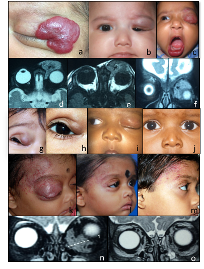
A thorough examination is necessary for capillary infantile hemangiomas and incorporates clinical assessment, imaging, and, in some situations, laboratory testing to verify the diagnosis, assess the lesion's extent, rule out consequences, and look for syndromic correlations. While clinical examination is the main method used to diagnose capillary infantile hemangiomas, additional diagnostic workup may be required in certain cases due to unusual presentations, deep lesions, or hemangiomas at key anatomical sites (see Image. Capillary Infantile Hemangiomas). The location, size, depth, and possible systemic involvement of the hemangioma determine which diagnostic instruments are used.[31]
Clinical Assessment A thorough clinical history and physical examination are the main components of the initial evaluation of capillary infantile hemangiomas. Clinicians should meticulously record the lesion's size, color, texture, and growth pattern. Important characteristics that assist in distinguishing capillary infantile hemangiomas from other vascular anomalies include the rate of proliferation, the depth of the lesion (superficial, deep, or mixed), and changes in size with weeping or physical activity. Furthermore, because of the potential for problems, lesions close to the eye, airway, or major blood vessels may need further investigation, making the location of the hemangioma crucial.[32]
A clinician examining a hemangioma needs to look for indications of functional impairment, including airway compromise (eg, in subglottic hemangiomas) or sight blockage (eg, periocular lesions). Systemic involvement indicators should be suspected in the presence of multiple cutaneous hemangiomas, which may indicate visceral lesions (eg, the liver).[19]
Imaging Indications Not every capillary infantile hemangioma necessitates imaging, particularly for minor, superficial lesions.[33][34] However, in the following situations, imaging studies become crucial:
- Deep or subcutaneous hemangiomas: These lesions can appear as lumps, discolorations, or swelling and may not be evident on the surface. Imaging can evaluate their depth, relationship to underlying structures, and potential complications, such as compressive effects on neighboring tissues.[35]
- Periocular hemangiomas: Hemangiomas near the eyes may result in visual axis obstruction, astigmatism, or amblyopia. Magnetic resonance imaging (MRI), in particular, is crucial to assess how the lesion affects the orbital structures and guarantee prompt intervention to protect eyesight.[36]
- Airway hemangiomas: Imaging is required to determine the size of the lesion and its impact on the airway lumen due to subglottic hemangiomas, which can cause airway obstruction. Any indications of respiratory distress, stridor, or hoarseness should prompt airway imaging. MRI or laryngoscopy are the usual methods used for this.[37]
- Large or segmental hemangiomas: PHACES syndrome may be linked to segmental hemangiomas, especially those on the face or neck. Imaging evaluates structural and vascular abnormalities, including intracranial malformations, cardiac defects, and arterial dysplasia. Furthermore, to rule out visceral involvement (eg, hepatic hemangiomas), individuals with numerous cutaneous lesions may benefit from liver ultrasonography.[38]
- Hemangiomas with rapid or atypical growth: Imaging may be necessary to distinguish rapidly expanding hemangiomas or lesions from other vascular anomalies such as congenital hemangiomas, rhabdomyosarcoma, or vascular malformations.[39]
- Complications: Especially in deeper tissue layers, signs of ulceration, infection, or bleeding may require imaging to determine the entire extent of the lesion.[40]
Imaging Modalities
Several diagnostic modalities may be utilized when evaluating apillary infantile hemangiomas.[17] Ultrasound
Ultrasound (US) imaging is the initial modality for assessing infant hemangiomas, especially subcutaneous and superficial lesions. Capillary infantile hemangiomas are usually seen on B-mode ultrasound as a well-demarcated, hypoechoic lesion. When evaluated with color Doppler ultrasound, the lesion frequently exhibits high vascularity, and the interior structure may look diverse. Increased blood flow is shown on Doppler US, supporting the diagnosis of a highly vascular lesion such as a hemangioma. This method is beneficial in evaluating the degree of vascularity and informing treatment choices (eg, figuring out how propranolol could work). US is also beneficial for serial monitoring of lesion size and response to treatment, especially when systemic therapies, eg, beta-blockers, are used.[41][41]Magnetic Resonance Imaging
MRI is the preferred imaging modality for assessing deep or complicated hemangiomas and those affecting important structures such as the orbit or airway. Due to the lesion's high water content and vascularity, capillary infantile hemangiomas usually appear on MRI as hyperintense on T2-weighted images and hypointense on T1-weighted images. Flow voids are a crucial feature of hemangiomas on MRIs; they show blood moving quickly through the tumor's vascular channels. MRI with gadolinium enhancement displays strong, homogenous enhancement, which aids in differentiating hemangiomas from other vascular abnormalities or soft tissue masses. MRI is particularly beneficial in determining the lesion's relationship to adjacent structures, such as the airway, blood vessels, or nerves. Magnetic resonance angiography (MRA) may be performed on patients with PHACES syndrome to look for cardiac anomalies, intracranial malformations, and arterial abnormalities.[42]
Computerized tomography
Because of the radiation exposure, computerized tomography (CT) is not frequently utilized for regular examination of IHs; however, in emergent cases, where quick imaging is required to check for problems such as bone erosion or airway obstruction, it can be helpful. On CT, hemangiomas appear as well-circumscribed soft tissue masses that could get brighter when contrast is injected intravenously. Their non-calcified characteristics aid in their differentiation from other lesions. When evaluating for related vascular abnormalities, CT angiography (CTA) is a useful diagnostic tool, particularly for patients who may have PHACES syndrome. However, MRI and MRA are typically chosen because they don't use ionizing radiation and have better soft-tissue resolution.[43][17][43] Laryngoscopy
Direct laryngoscopy or bronchoscopy is crucial for diagnosing suspected subglottic hemangiomas, especially where airway involvement is a concern. Laryngoscopy can identify submucosal hemangiomas that may not be apparent on external inspection and provide direct vision of the airway. Diagnosing these lesions, which frequently resemble bluish subglottic masses, is essential to preventing airway compromise.[44][17][44]
Angiography
Although infrequently required, angiography might be performed when vascular malformations or arteriovenous shunting are of concern, especially in situations with large, intricate hemangiomas. This imaging modality lets a clinician see the lesion's venous drainage and blood supply in great detail. Because MRI and CT angiography have advanced, Digital subtraction angiography (DSA) is used less often. Nevertheless, DSA is still useful when surgical intervention is planned or when embolization is necessary for complex hemangiomas.[45]
Laboratory Studies Since most cases of capillary infantile hemangiomas are predominantly clinical and supported by imaging, laboratory testing is usually unnecessary. Nonetheless, certain laboratory tests could be necessary in certain circumstances, including:
- Complete blood count: A complete blood count (CBC) is required to assess for thrombocytopenia and possible anemia owing to blood sequestration inside the hemangioma in patients with suspected Kasabach-Merritt syndrome, an uncommon complication involving thrombocytopenia and coagulopathy.
- Liver function tests: Hepatic hemangiomas are dangerous in newborns with multiple cutaneous hemangiomas, particularly if >5 lesions are present. To evaluate for hepatic involvement, liver function tests and abdominal ultrasound are frequently conducted. Further research is necessary because hepatic hemangiomas have the potential to cause high-output cardiac failure as a result of shunting in extreme situations.
- Thyroid function tests: In rare cases, hypothyroidism may result from the deiodination of thyroid hormone brought on by massive hepatic hemangiomas. Thyroid function tests, particularly in the liver, may be prescribed to screen for hypothyroidism in individuals with large or numerous hemangiomas.
- Genetic testing: While not always necessary, genetic testing may be suitable to assess for underlying chromosomal or genetic abnormalities in cases of syndromic hemangiomas or when genetic syndromes (eg, PHACES syndrome or CLOVES syndrome) are suspected. Genetic counseling and testing may also be necessary in these situations.
Treatment / Management
Capillary infantile hemangiomas are most often asymptomatic and thus require no intervention. About 10% of capillary infantile hemangiomas can present with astigmatism, visually obscuring ptosis, amblyopia, ulceration or bleeding, significant proptosis causing optic nerve compression, or exposure keratopathy. These complications require management. Treatment primarily involves beta-blockers, with oral propranolol and topical timolol as the mainstays. Other treatments, like lasers and surgery, are considered when beta-blockers are ineffective or complications persist. Close monitoring is essential to mitigate potential adverse effects.[46](A1)
Beta-Blocker Therapy
Topical or systemic beta-blockers are currently the mainstay for capillary infantile hemangioma management. Following their accidental discovery by Leaute-Labreze and colleagues in 2008, who noticed a regression in cutaneous hemangiomas in children treated with beta-blockers for cardiac and renal problems, propranolol has replaced oral steroids as the treatment modality of choice.[47](B3)
Beta-blockers suppress VEGF and fibroblast growth factors (FGF), which are responsible for proliferation. They also cause down-regulation of cyclic AMP needed for cell signaling and induce apoptosis of proliferating cells.[48] Treatment options using beta-blockers include:
- Oral propranolol: Propranolol is started at a low dosage of 0.16 mg/kg and gradually increased to 2 mg/kg/day in 3 doses without complications.[49] The medicine is usually taken for 6 to 12 months, depending on how well the patient responds to the treatment. The treatment period may be extended into the involution phase to stop rebound growth.
- Topical timolol: Timolol is applied twice daily as a 0.5% gel until the lesions regress and is helpful for superficial lesions.[50] When administered directly to the lesion, timolol, this nonselective beta-blocker akin to propranolol, reduces blood flow and encourages the involution of superficial hemangiomas locally. Timolol is very helpful for superficial, tiny hemangiomas that are bothersome from a cosmetic standpoint but not close to any critical structures. (B2)
A reduction in size and change in color can be noticed as early as 1 week after commencing treatment. Treatment should continue throughout the proliferative phase (up to 12 months of age) and gradually taper and stop to avoid a rebound increase in size. Some patients may experience adverse effects. Potential adverse effects include hypotension, bradycardia, hypoglycemia, bronchospasm, sleep disturbances, diarrhea, and hyperkalemia. These can be overcome by close monitoring during initial administration and proper parental counseling.
Corticosteroids
Before the advent of beta-blockers, corticosteroids were administered orally or intralesionally. This was the treatment of choice for capillary hemangiomas.[51] Steroids are effective only during the early proliferative phase and are associated with significant adverse effects. Prolonged use of corticosteroids is linked to several serious adverse effects that include cushingoid features, growth retardation, weight gain, adrenal suppression, hypertension, immune suppression, localized hypopigmentation of skin, and fat atrophy. Central retinal artery occlusion has been reported, thus making this modality a less preferred choice. Steroid injections into lesions that are being removed surgically are still used.[52]
Corticosteroids are currently regarded as a second-line therapy in cases when beta-blockers are inappropriate or do not work.[53] They decrease endothelial proliferation and inhibit angiogenesis. Like beta-blockers, they cause apoptosis in hemangioma cells, but their effects are more systemic. Oral prednisone, depending on the clinical response, is normally tapered over several weeks to months at a dose of 2 to 4 mg/kg/day. Intralesional corticosteroids (eg, betamethasone or triamcinolone) are used to minimize the size of the hemangioma before surgery, particularly in cases when surgical removal is anticipated.[31](A1)
Immunomodulators
Several immunomodulating agents have been tried in steroid-resistant cases. However, these agents are associated with complications such as bone marrow suppression and hepatotoxicity. Immunomodulators provide a focused method of treating capillary infantile hemangiomas, particularly in cases where other therapies are ineffective. In situations where propranolol or corticosteroids are inappropriate or ineffectual, they offer substitutes. Certain immunomodulators, such as sirolimus, have effectively treated complex hemangiomas linked to syndromic disorders or their consequences, such as ulceration and airway obstruction. Possible adverse effects, especially when using systemic immunomodulators like sirolimus and IFN-α, might impair immune response and raise the risk of infection. Concerns have been raised about the long-term safety of newborns due to the possibility of neurotoxicity and growth delays.[54](A1)
Interferon-alpha (IFN-α) works by downregulating pro-angiogenic factors, eg, FGF and VEGF.[55] IFN-α prevents angiogenesis, or the production of new blood vessels, which is essential to the growth of hemangiomas. IFN-α also boosts the immune system's capacity to control aberrant growth by promoting macrophage and natural killer cell activity. In the past, IFN-α was used to treat hemangiomas that were potentially fatal or functionally impaired, especially when previous treatments had failed. It is applied subcutaneously and takes a long time to work (usually several months). IFN-α can cause hemangiomas to shrink, especially those that are not responsive to corticosteroids or propranolol. Despite its usefulness in certain circumstances, its use has reduced due to concerns about major adverse effects, most notably neurotoxicity leading to spastic diplegia. Safer alternatives have mainly supplanted IFN-α because of the potential of neurodevelopmental adverse effects, such as spasticity and motor delays, which is the main limitation of its usage in babies. Sirolimus is an inhibitor of mTOR (mechanistic target of rapamycin) with antiangiogenic and immunosuppressive qualities. It obstructs the mTOR pathway, which is connected to angiogenesis, cell division, and growth.[56] By impeding this pathway, sirolimus lessens the aberrant vascular growth observed in hemangiomas. Complex infantile hemangiomas may benefit from sirolimus treatment, especially if they are resistant to propranolol or linked to conditions like PHACE syndrome. Treatment is given orally, and the length of time varies based on how severe the hemangioma is. Sirolimus has had encouraging outcomes in early clinical reports, particularly when treating troublesome hemangiomas because of ulceration, airway involvement, or rapid growth. Notable tumor reduction and symptom relief have been noted in some cases. Although sirolimus is usually well tolerated, it can weaken the immune system, raising the risk of infections. Other adverse effects include hyperlipidemia, gastrointestinal problems, and mouth ulcers.Imiquimod is an immunomodulator applied topically that promotes the synthesis of cytokines important in immunological regulation and antiviral responses, including interleukins, tumor necrosis factor-alpha (TNF-α), and IFN-α.[57] It might inhibit angiogenesis and encourage the hemangioma tissue to recede by strengthening the local immune response. Imiquimod is used topically and has been investigated for tiny or superficial hemangiomas, especially as a stand-alone treatment or in situations where other therapies are not appropriate. Some studies have suggested imiquimod may help reduce the size and color of superficial hemangiomas. However, the findings have been inconsistent. Usually, imiquimod is only used in minor cases that don't involve significant penetration. Common adverse effects include soreness, redness, and local skin irritation. Because of the topical administration, systemic absorption and extensive immunological modification are less of an issue.Lasers(A1)
Superficial hemangiomas can be treated with pulsed–dye lasers, which diminish the size and lighten the color of the lesions.[58] For superficial hemangiomas, especially those in the proliferative phase or residual telangiectasias following hemangioma involution, laser therapy, namely with pulsed-dye lasers (PDL),[59] is a viable therapeutic option. PDL selectively destroys aberrant blood vessels without endangering adjacent tissue by targeting the oxyhemoglobin in the hemangioma's blood vessels. As a result, the hemangioma lightens and shrines. Indications include superficial hemangiomas that do not show improvements with medication.[60] Ulcerated hemangiomas can be considered. Laser therapy can aid in healing by lowering inflammation and blood flow. Laser treatment can also address cosmetic concerns post-involution in cases where telangiectasias or other persistent vascular abnormalities persist. Difficult lesions may need multiple sessions for best results from PDL treatments, normally administered in an outpatient setting.[61] Mild erythema, edema, and crusting at the treatment site are common adverse effects that usually go away in a few days.[62] When used properly, there is very little chance of scarring. Surgery(A1)
When other treatments, such as medications (eg, beta-blockers like propranolol) or laser therapy, have failed or are deemed inappropriate, surgery for capillary infantile hemangiomas is typically considered.[63] In instances where the growths result in significant disfigurement or ulceration or if they impair vital functions, eg, vision or breathing, surgical intervention may be necessary,[64] even though most hemangiomas resolve spontaneously over time. The age and health of the infant, as well as the size, location, and severity of the hemangioma, all contribute to the decision to undergo surgery. Excision of the lesion is a technique that can lead to immediate removal, but it also entails risks, such as scarring or damage to the surrounding tissues.
Conjunctival capillary infantile hemangiomas are slow-growing tumors from endothelial cells. The best treatment option is an excisional biopsy, cauterization as they bleed torrentially, and closure of the conjunctiva as these lesions do not resolve with observation or medical management. Conjunctival capillary infantile hemangiomas can occur alone or as part of Sturge-Weber Syndrome.
Surgery is generally reserved for situations where the benefits outweigh the potential risks, and it is frequently regarded as a last resort option when more conservative treatments are ineffective. Surgical debulking of the lesion is reserved only for large vision-threatening lesions. As these lesions are irregular and unencapsulated, a complete removal is impossible. However, most large orbital and eyelid hemangiomas can be successfully treated with careful surgical dissection. Concurrent steroids are injected into any residual hemangioma tissue during surgery.[65]
Differential Diagnosis
The location and size of the lesion may be mistaken for other benign conditions, including lymphangiomas, arteriovenous malformations, mucoceles, or meningoceles. Lesions presenting with rapid-onset proptosis can mimic rhabdomyosarcoma or lymphangiomas. Pyogenic granuloma is a vascular lesion that usually develops after trauma and is sometimes mistaken for a hemangioma. Vascular malformations must also be considered in the differential diagnosis. These develop in line with the child's growth and are present from birth but don't have the proliferative phase typical of capillary infantile hemangiomas.
Staging
Since capillary infantile hemangiomas are benign lesions with a predictable course, they are not usually staged. Several studies have proposed staging systems based on location, size, and the occurrence of complications to help with treatment decisions. Hemangiomas near essential structures such as the eyes or airway could be categorized as high-risk and require more stringent management.
Prognosis
The overall prognosis for capillary infantile hemangiomas is excellent, as these lesions usually involute spontaneously. A more cautious prognosis may apply to complex diseases, eg, those involving the eye or airway, especially if they are not treated immediately. Large lesions respond very well to oral propranolol, which results in complete to the near-complete resolution of the lesions.[48]
Complications
Although most capillary infantile hemangiomas involute over time, some will undergo rapid growth and can cause amblyopia, proptosis with exposure keratitis, or optic nerve compression. Ulceration and bleeding may occur but are infrequently seen. Amblyopia can be brought on by hemangiomas blocking the visual axis (ie, hemangiomas of the eyelids). The ulceration of superficial hemangiomas can cause bleeding, discomfort, or infection. Life-threatening obstruction of the airway can result from large hemangiomas. Merritt-Kasabach Syndrome is a rare but dangerous side effect that is linked to big hemangiomas and is characterized by thrombocytopenia and coagulopathy.
Treatment can also present with side effects. Complications of oral steroids include hypotension, bradycardia, hypoglycemia, bronchospasm, sleep disturbances, diarrhea, and hyperkalemia. These can be overcome by close monitoring during initial administration and proper parental counseling. Complications of surgical resection include injury to the surrounding tissues, such as the levator aponeurosis and levator muscle. Orbital capillary hemangioma resection often results in a residual tumor in the orbit because these lesions are irregular with no capsulation.
Deterrence and Patient Education
Information regarding the natural course of infantile hemangiomas should be provided to parents. Most lesions will heal independently, but others will need medical intervention. Reminding parents of the natural history of capillary infantile hemangiomas is essential. With modern treatments using oral propranolol, most capillary infantile hemangiomas that require treatment will respond, although the procedure may need to be continued for a prolonged period. Complex lesions should be sent to a professional immediately. The safety and effectiveness of beta-blockers and the significance of monitoring for complications (eg, hypotension or hypoglycemia) should be explained to parents during treatment.
Pearls and Other Issues
Capillary infantile hemangiomas are typically benign lesions that do not cause complications. By treating periocular hemangiomas as soon as possible with beta-blockers, amblyopia can be avoided. Multidisciplinary care is essential for handling complex cases, especially those involving the system (eg, PHACES syndrome). Imaging is not always necessary, but it may be crucial in cases with deep or intricate hemangiomas.
Enhancing Healthcare Team Outcomes
Capillary infantile hemangiomas are common benign lesions, and parents can present to either pediatricians, ophthalmologists, oculoplastic surgeons, or dermatologists with complaints of reddish discoloration of the skin or as an eyelid mass. Healthcare physicians should be aware of this common clinical entity and its features, enabling them to distinguish it from other more severe soft tissue lesions present during childhood. Early treatment with oral propranolol in coordination with a pediatrician shows excellent results. Careful attention should be paid to assessing refraction and vision to detect astigmatism, refractive error, or amblyopia. Sequential imaging may be necessary in the presence of proptosis due to an intraorbital hemangioma. The cornea should be carefully examined for any evidence of exposure keratopathy.
An interprofessional team comprising pediatricians, dermatologists, ophthalmologists, and surgeons should manage complicated capillary infantile hemangiomas. Coordinated care guarantees prompt intervention for patients with complex lesions, like those affecting the eyes or airway. A strategic approach is equally crucial, involving evidence-based strategies to optimize treatment plans and minimize adverse effects. When beta-blockers are provided, close monitoring for adverse effects (eg, hypotension, bradycardia) is required. Therefore, regular communication between specialists is essential to achieving the best results. Collaborative decision-making with the family promotes adherence to long-term treatment programs and helps to set expectations.
Media
(Click Image to Enlarge)
Capillary Infantile Hemangiomas. Image A: Clinical picture showing a large superficial hemangioma. Image B, C: Partial and complete obscuration of visual axis due to hemangioma of the upper lid. Images D, E, F: MRI scans axial and coronal view showing homogenous solid preseptal and postseptal soft tissue lesion, which is isointense on T1W1 and hyperintense on T2W2 with characteristic flow voids within. Images G, H, I, J: Clinical pictures showing partial and complete resolution of upper lid hemangioma with oral propranolol therapy. Images K, L, M: Clinical picture showing hemangioma at presentation and a significant reduction in size and color of the lesion at 3 months and 12 months posttreatment with oral propranolol. Images N, O: Pretreatment and posttreatment MRI scan showing complete resolution of lesion located along the medial and inferior extraconal space with oral propranolol.
Contributed by BCK Patel, MD, FRCS, and K Koka, MD
References
Spiteri Cornish K, Reddy AR. The use of propranolol in the management of periocular capillary haemangioma--a systematic review. Eye (London, England). 2011 Oct:25(10):1277-83. doi: 10.1038/eye.2011.164. Epub 2011 Jul 8 [PubMed PMID: 21738233]
Level 1 (high-level) evidenceMulliken JB, Glowacki J. Hemangiomas and vascular malformations in infants and children: a classification based on endothelial characteristics. Plastic and reconstructive surgery. 1982 Mar:69(3):412-22 [PubMed PMID: 7063565]
Gupta R, Bhandari A, Navarro OM. Pediatric Vascular Anomalies: A Clinical and Radiological Perspective. The Indian journal of radiology & imaging. 2024 Jan:34(1):103-127. doi: 10.1055/s-0043-1774391. Epub 2023 Sep 16 [PubMed PMID: 38106867]
Level 3 (low-level) evidenceSatterfield KR, Chambers CB. Current treatment and management of infantile hemangiomas. Survey of ophthalmology. 2019 Sep-Oct:64(5):608-618. doi: 10.1016/j.survophthal.2019.02.005. Epub 2019 Feb 14 [PubMed PMID: 30772366]
Level 3 (low-level) evidenceNeri I, Orioni G, Leuzzi M, Facchini E, Mengozzi E, Chessa MA. Infantile Hemangioma with Minimal or Arrested Growth (IHMAG): A Retrospective Analysis of Clinical and Dermoscopic Diagnostic Clues. Dermatology practical & conceptual. 2024 Jul 1:14(3):. doi: 10.5826/dpc.1403a206. Epub 2024 Jul 1 [PubMed PMID: 39122516]
Level 2 (mid-level) evidenceSơn TT, Nghĩa PT, Dung PTV, Thuý TTH. Reconstructive techniques for radiation-induced hemifacial sequelae: A hemangioma case study. International journal of surgery case reports. 2024 Jul:120():109906. doi: 10.1016/j.ijscr.2024.109906. Epub 2024 Jun 12 [PubMed PMID: 38880001]
Level 3 (low-level) evidenceMahajan P, Bergstrom KL, Phung TL, Metry DW. The genetics of vascular birthmarks. Clinics in dermatology. 2022 Jul-Aug:40(4):313-321. doi: 10.1016/j.clindermatol.2022.02.006. Epub 2022 Feb 16 [PubMed PMID: 35181412]
Smith CJF, Friedlander SF, Guma M, Kavanaugh A, Chambers CD. Infantile Hemangiomas: An Updated Review on Risk Factors, Pathogenesis, and Treatment. Birth defects research. 2017 Jul 3:109(11):809-815. doi: 10.1002/bdr2.1023. Epub 2017 Apr 12 [PubMed PMID: 28402073]
Oszajca K, Szemraj J, Wyrzykowski D, Chrzanowska B, Salamon A, Przewratil P. Single-nucleotide polymorphisms of VEGF-A and VEGFR-2 genes and risk of infantile hemangioma. International journal of dermatology. 2018 Oct:57(10):1201-1207. doi: 10.1111/ijd.14127. Epub 2018 Jul 9 [PubMed PMID: 29984822]
Dickison P, Christou E, Wargon O. A prospective study of infantile hemangiomas with a focus on incidence and risk factors. Pediatric dermatology. 2011 Nov-Dec:28(6):663-669. doi: 10.1111/j.1525-1470.2011.01568.x. Epub 2011 Oct 13 [PubMed PMID: 21995808]
Tiple S, Kimmatkar P, Das S, Muralidhara A, Nehashree, Mehta A, Patidar N, Jain E. Treatment outcomes of oral propranolol in the treatment of periocular infantile capillary hemangioma and factors predictive of recurrence and incomplete resolution: A multi-centric study. Oman journal of ophthalmology. 2023 Jan-Apr:16(1):75-81. doi: 10.4103/ojo.ojo_11_22. Epub 2023 Feb 21 [PubMed PMID: 37007245]
Sun Z, Li M, Dong C, Mei S. Clinical characteristics of infantile haemangioma in twins: a retrospective study. BMC pediatrics. 2024 Feb 13:24(1):111. doi: 10.1186/s12887-024-04602-8. Epub 2024 Feb 13 [PubMed PMID: 38350912]
Level 2 (mid-level) evidenceChen XD, Ma G, Chen H, Ye XX, Jin YB, Lin XX. Maternal and perinatal risk factors for infantile hemangioma: a case-control study. Pediatric dermatology. 2013 Jul-Aug:30(4):457-61. doi: 10.1111/pde.12042. Epub 2012 Dec 26 [PubMed PMID: 23278441]
Level 2 (mid-level) evidenceTurhan AB, Bör Ö, Özdemir ZC. Treatment with propranolol for infantile hemangiomas: single-center experience. Journal of cosmetic dermatology. 2016 Sep:15(3):296-302. doi: 10.1111/jocd.12220. Epub 2016 Apr 7 [PubMed PMID: 27061310]
Munden A, Butschek R, Tom WL, Marshall JS, Poeltler DM, Krohne SE, Alió AB, Ritter M, Friedlander DF, Catanzarite V, Mendoza A, Smith L, Friedlander M, Friedlander SF. Prospective study of infantile haemangiomas: incidence, clinical characteristics and association with placental anomalies. The British journal of dermatology. 2014 Apr:170(4):907-13. doi: 10.1111/bjd.12804. Epub [PubMed PMID: 24641194]
Amrock SM, Weitzman M. Diverging racial trends in neonatal infantile hemangioma diagnoses, 1979-2006. Pediatric dermatology. 2013 Jul-Aug:30(4):493-4. doi: 10.1111/pde.12075. Epub 2013 Jan 17 [PubMed PMID: 23331036]
Haik BG, Karcioglu ZA, Gordon RA, Pechous BP. Capillary hemangioma (infantile periocular hemangioma). Survey of ophthalmology. 1994 Mar-Apr:38(5):399-426 [PubMed PMID: 8009426]
Level 3 (low-level) evidenceCouto RA, Maclellan RA, Zurakowski D, Greene AK. Infantile hemangioma: clinical assessment of the involuting phase and implications for management. Plastic and reconstructive surgery. 2012 Sep:130(3):619-624. doi: 10.1097/PRS.0b013e31825dc129. Epub [PubMed PMID: 22575857]
Rodríguez Bandera AI, Sebaratnam DF, Wargon O, Wong LF. Infantile hemangioma. Part 1: Epidemiology, pathogenesis, clinical presentation and assessment. Journal of the American Academy of Dermatology. 2021 Dec:85(6):1379-1392. doi: 10.1016/j.jaad.2021.08.019. Epub 2021 Aug 19 [PubMed PMID: 34419524]
Hasbani DJ, Hamie L. Infantile Hemangiomas. Dermatologic clinics. 2022 Oct:40(4):383-392. doi: 10.1016/j.det.2022.06.004. Epub 2022 Sep 16 [PubMed PMID: 36243426]
Sun Y, Qiu F, Hu C, Guo Y, Lei S. Hemangioma Endothelial Cells and Hemangioma Stem Cells in Infantile Hemangioma. Annals of plastic surgery. 2022 Feb 1:88(2):244-249. doi: 10.1097/SAP.0000000000002835. Epub [PubMed PMID: 35023872]
Level 2 (mid-level) evidenceFagiani E, Lorentz P, Bill R, Pavotbawan K, Kopfstein L, Christofori G. VEGF receptor-2-specific signaling mediated by VEGF-E induces hemangioma-like lesions in normal and in malignant tissue. Angiogenesis. 2016 Jul:19(3):339-58. doi: 10.1007/s10456-016-9508-7. Epub 2016 Apr 1 [PubMed PMID: 27038485]
Höger PH, Hamm H. [Infantile hemangioma : Clinical manifestation, treatment, and differential diagnoses]. Dermatologie (Heidelberg, Germany). 2023 May:74(5):331-339. doi: 10.1007/s00105-023-05145-2. Epub [PubMed PMID: 37083897]
Godar M, Yuan Q, Shakya R, Xia Y, Zhang P. Mixed capillary venous retroperitoneal hemangioma. Case reports in radiology. 2013:2013():258352. doi: 10.1155/2013/258352. Epub 2013 Feb 28 [PubMed PMID: 23533905]
Level 3 (low-level) evidenceJakobiec FA, Hanbazazh M, Cortes Barrantes P, Chodosh J. Adult Primary Capillary Hemangioma of the Sclera: A Previously Undescribed Entity With a Review of Epibulbar Vascular Lesions. Ophthalmic plastic and reconstructive surgery. 2020 Jul/Aug:36(4):327-333. doi: 10.1097/IOP.0000000000001582. Epub [PubMed PMID: 31895731]
Lan J, Huang B, Liu R, Ju X, Zhou Y, Jiang J, Liang W, Shen Y, Li F, Pang L. Expression of cancer stem cell markers and their correlation with pathogenesis in vascular tumors. International journal of clinical and experimental pathology. 2015:8(10):12621-33 [PubMed PMID: 26722452]
van Vugt LJ, van der Vleuten CJM, Flucke U, Blokx WAM. The utility of GLUT1 as a diagnostic marker in cutaneous vascular anomalies: A review of literature and recommendations for daily practice. Pathology, research and practice. 2017 Jun:213(6):591-597. doi: 10.1016/j.prp.2017.04.023. Epub 2017 Apr 27 [PubMed PMID: 28552538]
Stass-Isern M. Periorbital and orbital infantile hemangiomas. International ophthalmology clinics. 2014 Summer:54(3):73-82. doi: 10.1097/IIO.0000000000000039. Epub [PubMed PMID: 24879105]
Osman NM. Kasabach - Merritt syndrome: A case report. Sudanese journal of paediatrics. 2013:13(1):49-52 [PubMed PMID: 27493358]
Level 3 (low-level) evidenceFrieden IJ, Reese V, Cohen D. PHACE syndrome. The association of posterior fossa brain malformations, hemangiomas, arterial anomalies, coarctation of the aorta and cardiac defects, and eye abnormalities. Archives of dermatology. 1996 Mar:132(3):307-11 [PubMed PMID: 8607636]
Level 3 (low-level) evidenceSebaratnam DF, Rodríguez Bandera AL, Wong LF, Wargon O. Infantile hemangioma. Part 2: Management. Journal of the American Academy of Dermatology. 2021 Dec:85(6):1395-1404. doi: 10.1016/j.jaad.2021.08.020. Epub 2021 Aug 19 [PubMed PMID: 34419523]
Mulinda C, Yun S, Fenner J, Garzon MC, Scollan ME, Levin LE. How many is too many? A review of the significant numbers in pediatric skin lesions and their recommended evaluation. Pediatric dermatology. 2024 Sep 5:():. doi: 10.1111/pde.15687. Epub 2024 Sep 5 [PubMed PMID: 39235117]
Levin LE, Lauren CT. Multifocal vascular lesions. Seminars in cutaneous medicine and surgery. 2016 Sep:35(3):153-60. doi: 10.12788/j.sder.2016.054. Epub [PubMed PMID: 27607324]
Azizkhan RG. Complex vascular anomalies. Pediatric surgery international. 2013 Oct:29(10):1023-38. doi: 10.1007/s00383-013-3393-6. Epub [PubMed PMID: 23989523]
Piccolo V, Russo T, Moscarella E, Brancaccio G, Alfano R, Argenziano G. Dermatoscopy of Vascular Lesions. Dermatologic clinics. 2018 Oct:36(4):389-395. doi: 10.1016/j.det.2018.05.006. Epub 2018 Jul 31 [PubMed PMID: 30201148]
Spence-Shishido AA, Good WV, Baselga E, Frieden IJ. Hemangiomas and the eye. Clinics in dermatology. 2015 Mar-Apr:33(2):170-82. doi: 10.1016/j.clindermatol.2014.10.009. Epub [PubMed PMID: 25704937]
Kreher MA, Siegel LH, Shmuylovich L. A case of large airway and orbital hemangiomas with facial capillary malformation. Pediatric dermatology. 2023 Nov-Dec:40(6):1142-1144. doi: 10.1111/pde.15340. Epub 2023 May 10 [PubMed PMID: 37164359]
Level 3 (low-level) evidenceHeyer GL. PHACE(S) syndrome. Handbook of clinical neurology. 2015:132():169-83. doi: 10.1016/B978-0-444-62702-5.00012-3. Epub [PubMed PMID: 26564079]
Diociaiuti A, Paolantonio G, Zama M, Alaggio R, Carnevale C, Conforti A, Cesario C, Dentici ML, Buonuomo PS, Rollo M, El Hachem M. Vascular Birthmarks as a Clue for Complex and Syndromic Vascular Anomalies. Frontiers in pediatrics. 2021:9():730393. doi: 10.3389/fped.2021.730393. Epub 2021 Oct 7 [PubMed PMID: 34692608]
Gautam A, Frieden IJ, Shah SD, Witman PM, Harfmann K, Bradley F, Blei F, Pope E, Alsumait A, Gupta D, Covelli I, Streicher JL, Cotton C, Tollefson M, Nguyen H, Hunt R, Buros A, Fernández Faith E. Clinical characteristics of infantile hemangiomas with aggressive, persistent, and destructive ulceration. Pediatric dermatology. 2023 Nov-Dec:40(6):996-1002. doi: 10.1111/pde.15444. Epub 2023 Oct 16 [PubMed PMID: 37845188]
Babic V, Schoeffler A, Moawad S, Mainard L, Schmutz JL, Bursztejn AC. [Rare presentations of infantile hemangiomas: 4 cases]. Annales de dermatologie et de venereologie. 2018 Dec:145(12):749-755. doi: 10.1016/j.annder.2018.07.026. Epub 2018 Nov 9 [PubMed PMID: 30415930]
Level 3 (low-level) evidenceFriedland R, Ben Amitai D, Zvulunov A. Screening for Brain Involvement in Infants with Multifocal Cutaneous Infantile Hemangiomas. Dermatology (Basel, Switzerland). 2017:233(6):435-440. doi: 10.1159/000484598. Epub 2018 Jan 20 [PubMed PMID: 29353270]
MacDougall MS, Afzal SY, Freedman MS, Han P. Infant in extremis: respiratory failure secondary to lower airway infantile hemangioma. BMC pediatrics. 2022 Dec 30:22(1):744. doi: 10.1186/s12887-022-03821-1. Epub 2022 Dec 30 [PubMed PMID: 36581920]
Parhizkar N, Manning SC, Inglis AF Jr, Finn LS, Chen EY, Perkins JA. How airway venous malformations differ from airway infantile hemangiomas. Archives of otolaryngology--head & neck surgery. 2011 Apr:137(4):352-7. doi: 10.1001/archoto.2010.243. Epub 2011 Jan 17 [PubMed PMID: 21242531]
Benzar I. A Diagnostic Program of Vascular Tumor and Vascular Malformations in Children According to Modern Classification. Acta medica (Hradec Kralove). 2017:60(1):19-26. doi: 10.14712/18059694.2017.47. Epub 2017 May 26 [PubMed PMID: 28550689]
Buckmiller LM, Munson PD, Dyamenahalli U, Dai Y, Richter GT. Propranolol for infantile hemangiomas: early experience at a tertiary vascular anomalies center. The Laryngoscope. 2010 Apr:120(4):676-81. doi: 10.1002/lary.20807. Epub [PubMed PMID: 20112413]
Level 1 (high-level) evidenceLéauté-Labrèze C, Dumas de la Roque E, Hubiche T, Boralevi F, Thambo JB, Taïeb A. Propranolol for severe hemangiomas of infancy. The New England journal of medicine. 2008 Jun 12:358(24):2649-51. doi: 10.1056/NEJMc0708819. Epub [PubMed PMID: 18550886]
Level 3 (low-level) evidenceKoka K, Mukherjee B, Agarkar S. Effect of oral propranolol on periocular Capillary Hemangiomas of Infancy. Pediatrics and neonatology. 2018 Aug:59(4):390-396. doi: 10.1016/j.pedneo.2017.11.021. Epub 2017 Dec 6 [PubMed PMID: 29301720]
Pandey V, Bera RN, Diwedi AND, Singh OP, Tiwari P. Color Doppler Ultrasound Indices as Predictors of Propranolol Response in Infantile Hemangioma: A Prospective Study. Indian pediatrics. 2024 Mar 15:61(3):237-242 [PubMed PMID: 38469839]
Yuan L, Wang J. Efficacy and Safety of Introduction Timolol Maleate by Manometer Microneedles among Infantile Hemangioma: A Retrospective Study in China. European journal of pediatric surgery : official journal of Austrian Association of Pediatric Surgery ... [et al] = Zeitschrift fur Kinderchirurgie. 2024 Jun:34(3):261-266. doi: 10.1055/s-0043-57238. Epub 2023 May 1 [PubMed PMID: 37127023]
Level 2 (mid-level) evidenceAletaha M, Salour H, Bagheri A, Raffati N, Amouhashemi N. Oral propranolol for treatment of pediatric capillary hemangiomas. Journal of ophthalmic & vision research. 2012 Apr:7(2):130-3 [PubMed PMID: 23275821]
Alsmman AH, Mounir A. Combined oral propranolol with intralesional injection of triamcinolone acetonide in treatment of infantile periocular hemangiomas. Clinical ophthalmology (Auckland, N.Z.). 2017:11():2177-2181. doi: 10.2147/OPTH.S153121. Epub 2017 Dec 8 [PubMed PMID: 29263646]
Li LL, Yan L, Luo MX, Cai YX, Mi XB, Zhang TD. Propranolol in the Treatment of Infantile Hemangiomas: A Meta-Analysis in Chinese Patients. Pediatric dermatology. 2015 Sep-Oct:32(5):e210-4. doi: 10.1111/pde.12627. Epub 2015 Jun 10 [PubMed PMID: 26060892]
Level 1 (high-level) evidenceHogeling M, Adams S, Wargon O. A randomized controlled trial of propranolol for infantile hemangiomas. Pediatrics. 2011 Aug:128(2):e259-66. doi: 10.1542/peds.2010-0029. Epub 2011 Jul 25 [PubMed PMID: 21788220]
Level 1 (high-level) evidenceSgonc R, Fuerhapter C, Boeck G, Swerlick R, Fritsch P, Sepp N. Induction of apoptosis in human dermal microvascular endothelial cells and infantile hemangiomas by interferon-alpha. International archives of allergy and immunology. 1998 Nov:117(3):209-14 [PubMed PMID: 9831809]
Kleinman EP, Blei F, Adams D, Greenberger S. Sirolimus for diffuse intestinal infantile hemangioma with PHACE features: systematic review. Pediatric research. 2023 May:93(6):1470-1479. doi: 10.1038/s41390-022-02325-z. Epub 2022 Sep 30 [PubMed PMID: 36180586]
Level 1 (high-level) evidenceHu L, Huang HZ, Li X, Lin XX, Li W. Open-label nonrandomized left-right comparison of imiquimod 5% ointment and timolol maleate 0.5% eye drops in the treatment of proliferating superficial infantile hemangioma. Dermatology (Basel, Switzerland). 2015:230(2):150-5. doi: 10.1159/000369164. Epub 2015 Jan 24 [PubMed PMID: 25633200]
Level 1 (high-level) evidenceBrauer JA, Geronemus RG. Laser treatment in the management of infantile hemangiomas and capillary vascular malformations. Techniques in vascular and interventional radiology. 2013 Mar:16(1):51-4. doi: 10.1053/j.tvir.2013.01.007. Epub [PubMed PMID: 23499132]
Sugimoto A, Aoki R, Toyohara E, Ogawa R. Infantile Hemangiomas Cleared by Combined Therapy With Pulsed Dye Laser and Propranolol. Dermatologic surgery : official publication for American Society for Dermatologic Surgery [et al.]. 2021 Aug 1:47(8):1052-1057. doi: 10.1097/DSS.0000000000003018. Epub [PubMed PMID: 34397540]
Chinnadurai S, Sathe NA, Surawicz T. Laser treatment of infantile hemangioma: A systematic review. Lasers in surgery and medicine. 2016 Mar:48(3):221-33. doi: 10.1002/lsm.22455. Epub 2015 Dec 29 [PubMed PMID: 26711436]
Level 1 (high-level) evidenceNakazono M, Kagimoto S, Koike T, Satake T, Maegawa J. Clinical Outcomes of Small Infantile Hemangiomas Treated With Pulsed Dye Laser. Dermatologic surgery : official publication for American Society for Dermatologic Surgery [et al.]. 2022 Aug 1:48(8):833-837. doi: 10.1097/DSS.0000000000003491. Epub 2022 May 17 [PubMed PMID: 35580249]
Level 2 (mid-level) evidenceLin L, Guo P, Cao Y, Li Q, Zhang J, Huo R. Combination of Sclerotherapy and Dual-Wavelength Laser in the Management of Infantile Hemangiomas in Chinese Infants. Dermatologic surgery : official publication for American Society for Dermatologic Surgery [et al.]. 2019 Oct:45(10):1253-1259. doi: 10.1097/DSS.0000000000001898. Epub [PubMed PMID: 30882500]
Menapace D, Mitkov M, Towbin R, Hogeling M. The changing face of complicated infantile hemangioma treatment. Pediatric radiology. 2016 Oct:46(11):1494-506. doi: 10.1007/s00247-016-3643-6. Epub 2016 Jul 23 [PubMed PMID: 27450406]
Men CJ, Ediriwickrema LS, Paik JS, Murdock J, Yen MT, Ng JD, Liu CY, Korn BS, Kikkawa DO. Surgical Intervention of Periocular Infantile Hemangiomas in the Era of β-Blockers. Ophthalmic plastic and reconstructive surgery. 2020 Jan/Feb:36(1):70-73. doi: 10.1097/IOP.0000000000001466. Epub [PubMed PMID: 31809485]
Hochman M. Timing and Rationale of Treatment: Achieving the "Best Result". Dermatologic clinics. 2022 Oct:40(4):379-382. doi: 10.1016/j.det.2022.06.003. Epub 2022 Sep 16 [PubMed PMID: 36243425]