Introduction
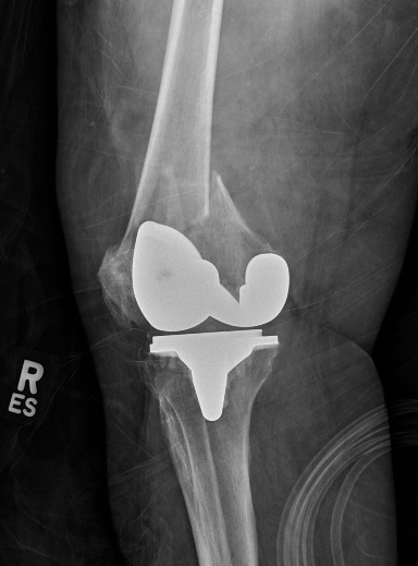
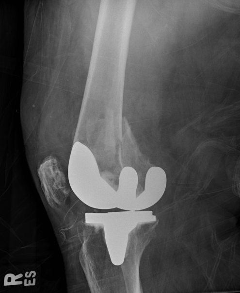
Knee arthroplasty is a reconstruction of the knee joint. It is more commonly referred to as a total knee replacement and is a very reliable procedure with predictable results. Total knee arthroplasty (TKA) is an excellent treatment option for individuals with symptomatic osteoarthritis in at least 2 of the 3 compartments of the knee and who have failed conservative treatment.[1][2] Additionally, partial knee arthroplasty (PKA) is an excellent treatment option for individuals with symptomatic osteoarthritis localized to 1 compartment of the knee and who have failed conservative treatment.[3] The primary goal of either surgery is durable pain relief with the improvement of functional status.
The history of TKA dates back to the mid to late 1800s when the first implants were made from ivory and fixed to the bone using a combination of colophony and plaster of Paris. This design did not produce favorable results and was eventually replaced with metal implants in the 1930s. In the 1950s, a hinged prosthesis was designed to replace the femur and tibia, as well as the stabilizing ligaments surrounding the knee. While this produced satisfactory results, there was a high rate of failure and poor long-term outcome due to the failure of reproducing the natural kinematics of the knee joint. This was subsequently replaced with a prosthesis that replicated the shape of the distal femur, preserved the collateral and cruciate ligaments, and consisted of a plastic bearing on the tibia. Since this breakthrough in prosthesis in the 1970s, the design has evolved to focus on replicating the anatomy and normal function of the knee joint. Furthermore, advances have been made in fixation techniques and wear properties of the bearing surface which positively affects the longevity of the knee replacement.[4]
Anatomy and Physiology
Register For Free And Read The Full Article
Search engine and full access to all medical articles
10 free questions in your specialty
Free CME/CE Activities
Free daily question in your email
Save favorite articles to your dashboard
Emails offering discounts
Learn more about a Subscription to StatPearls Point-of-Care
Anatomy and Physiology
The knee is a synovial hinge joint with minimal rotational motion. It is comprised of the distal femur, proximal tibia, and the patella. There are 3 separate articulations and compartments: medial femorotibial, lateral femorotibial, and patellofemoral. The stability of the knee joint is provided by the congruity of the joint as well as by the collateral ligaments. The capsule surrounds the entire joint and extends proximally into the suprapatellar pouch. Articular cartilage covers the femoral condyles, tibial plateaus, trochlear groove, and patellar facets. Menisci are interposed in the medial and lateral compartments between the femur and tibia which act to protect the articular cartilage and support the knee.
The mechanical axis of the femur, defined by a line drawn from the center of the femoral head to the center of the knee, is 3 degrees valgus to the vertical axis. The anatomic axis of the femur, defined by a line bisecting the femoral shaft, is 6 degrees valgus to the mechanical axis of the femur and 9 degrees valgus to the vertical axis.[5] The proximal tibia is oriented to 3 degrees of varus. The varus position of the proximal tibia, along with the offset of the hip center of rotation, results in the weight-bearing surface of the tibia being parallel to the ground. The sagittal alignment of the proximal tibia is sloped posteriorly approximately 5 to 7 degrees. The asymmetry of the natural bony anatomy maintains the alignment of the joint and ligamentous tension.
Indications
TKA is a well-described treatment option for patients suffering from knee pain secondary to osteoarthritis who have failed conservative treatment measures. It is a reliable procedure that provides pain relief and improves the patient’s functional status.[2] Furthermore, the need for correction of a significant or progressive deformity at the knee with evidence of osteoarthritis can also be an indication for a TKA. A patient with persistent knee pain without radiographic evidence of knee osteoarthritis should have further workup to exclude other possible sources of their pain.
Clinical symptoms of osteoarthritis include:
- Knee pain
- Pain with activity and improving with rest
- Pain gradually worsens over time
- Decreased ambulatory capacity
Clinical evaluation includes:
- Full knee exam including range of motion and ligamentous testing
- Knee radiographs include standing anteroposterior, lateral, 45-degree posteroanterior, and skyline view of the patella
Radiographic evidence of osteoarthritis include[6]:
- Joint space narrowing
- Subchondral sclerosis
- Subchondral cysts
- Osteophyte formation
Conservative treatment includes:
- Non-steroidal anti-inflammatory medication
- Weight loss
- Activity modification
- Bracing
- Physical therapy
- Viscosupplementation
- Intra-articular steroid injection
Contraindications
Absolute
- Active or latent (less than 1 year) knee sepsis
- Presence of active infection elsewhere in body
- Extensor mechanism dysfunction
- Medically unstable patient
Relative
- Neuropathic joint
- Poor overlying skin condition
- Morbid obesity
- Noncompliance due to major psychiatric disorder, alcohol, or drug abuse
- Insufficient bone stock for reconstruction
- Poor patient motivation or unrealistic expectation
- Severe peripheral vascular disease
Equipment
A TKA system will consist of instrumentation that helps the surgeon prepare the ends of the femur, tibia, and patella to receive an implant. The instrumentation will be specific to the brand and type of implant being used with each company and model having specific intricacies.
In general, the instrumentation will consist of:
- Intramedullary femoral guide to help establish the distal femoral alignment
- Distal femoral cutting guide
- Femoral sizing guide
- 4-in-1 femoral cutting guide
- Extramedullary or intramedullary tibial guide
- Proximal tibial cutting guide
- Patella sizing guide
- Femoral component trial
- Tibial baseplate trial
- Patellar button trial
- Trial plastic bearing
The final implants will come in individual sterile packages and will consist of:
- Femoral component, typically made of cobalt-chrome
- Tibial component, typically made of cobalt-chrome or titanium
- Tibial polyethylene bearing, made of an ultra high molecular weight (UHMW) polyethylene
- Patellar button, made of UHMW polyethylene
Personnel
- Anesthesia team
- Operating room nurse
- Surgical technician
- Surgical assistant
Preparation
- Full medical and drug history before surgery
- Appropriate pre-surgical workup, clearance, and optimization
- Pre-operative radiographs of the affected knee
- Pre-operative templating of the affected knee to estimate the component size
- Primary TKA system of choice
- Have various final implant sizes ready and available in the hospital
- Have increasing prosthesis constraint options ready and available in the hospital
- Have revision total knee replacement system of choice ready and available if needed
- +/- antibiotic cement, surgeon preference
Technique or Treatment
The goal of TKA is the same regardless of surgeon, implant, or technique. The variability in the procedure lies in the technique. Some of the variations in operative technique for TKA are listed below.
- General anesthesia versus regional anesthesia
- Tourniquet versus tourniquet-less surgery
- Standard versus patient-specific instrumentation
- Standard versus patient-specific implants
- Traditional versus robotic-assisted TKA
- Traditional versus navigation-assisted TKA
- Traditional versus sensor-assisted TKA
- Measured resection versus gap balancing
- Cruciate-retaining implant versus cruciate stabilized the implant
- Resurfaced versus non-resurfaced patella
- Cement versus cement-less (press fit) TKA
Complications
Potential complications include:[7][8]
- Infection, superficial and deep
- Blood clot
- Pulmonary embolism
- Fracture
- Dislocation
- Instability
- Osteolysis resulting in component loosening
- Pain
- Stiffness
- Vascular injury
- Nerve injury
Clinical Significance
The number of patients suffering from knee pain secondary to osteoarthritis will continue to rise, especially as the average life expectancy and obesity rates increase. These 2 factors contribute to the wear and tear of articular cartilage in the major weight-bearing joints seen in primary osteoarthritis.[9] Patients can also suffer from secondary osteoarthritis, or osteoarthritis caused by an abnormal concentration of force across the joint such as in rheumatoid or post-traumatic cases. In either case, a thorough history and physical and appropriate radiographs are essential to the appropriate diagnosis. Initial treatment is conservative and includes any and all combinations listed above. Once conservative treatment is no longer effective, surgical intervention may be considered. TKA is a reliable surgical procedure with a predictable outcome in the appropriate patient. Reported survival rates are upward of 85% with 10 to 25 years of follow-up.[10][11][10] Improvements in pain scores and functional scores are commonly recognized with the procedure as well.
Enhancing Healthcare Team Outcomes
While knee arthroplasty is done by the orthopedic surgeon, to ensure good outcomes an interprofessional team is required that includes a physical therapist, nurses, an internist, and the primary care provider. After surgery, the patients require physical therapy to regain muscle mass and joint function. In addition, for good outcomes, the patients need to exercise and maintain a healthy weight. Nurses should educate the patient on a healthy diet, avoidance of tobacco and taking part in regular exercise.
Media
(Click Image to Enlarge)
(Click Image to Enlarge)
References
Davies PS, Graham SM, Maqungo S, Harrison WJ. Total joint replacement in sub-Saharan Africa: a systematic review. Tropical doctor. 2019 Apr:49(2):120-128. doi: 10.1177/0049475518822239. Epub 2019 Jan 12 [PubMed PMID: 30636518]
Level 1 (high-level) evidenceAdie S, Harris I, Chuan A, Lewis P, Naylor JM. Selecting and optimising patients for total knee arthroplasty. The Medical journal of Australia. 2019 Feb:210(3):135-141. doi: 10.5694/mja2.12109. Epub 2019 Jan 18 [PubMed PMID: 30656689]
Naziri Q, Mixa PJ, Murray DP, Abraham R, Zikria BA, Sastry A, Patel PD. Robotic-Assisted and Computer-Navigated Unicompartmental Knee Arthroplasties: A Systematic Review. Surgical technology international. 2018 Jun 1:32():271-278 [PubMed PMID: 29611157]
Level 1 (high-level) evidenceStilling M, Mechlenburg I, Jepsen CF, Rømer L, Rahbek O, Søballe K, Madsen F. Superior fixation and less periprosthetic stress-shielding of tibial components with a finned stem versus an I-beam block stem: a randomized RSA and DXA study with minimum 5 years' follow-up. Acta orthopaedica. 2019 Apr:90(2):165-171. doi: 10.1080/17453674.2019.1566510. Epub 2019 Jan 23 [PubMed PMID: 30669918]
Level 1 (high-level) evidenceWang Z, Cheng X, Zhang Y, Zhang X, Zhou Y. Restoration of Constitutional Alignment in TKA with a Novel Osteotomy Technique. The journal of knee surgery. 2020 Feb:33(2):190-199. doi: 10.1055/s-0038-1677508. Epub 2019 Jan 16 [PubMed PMID: 30650441]
Minciullo L, Parkes MJ, Felson DT, Cootes TF. Comparing image analysis approaches versus expert readers: the relation of knee radiograph features to knee pain. Annals of the rheumatic diseases. 2018 Nov:77(11):1606-1609. doi: 10.1136/annrheumdis-2018-213492. Epub 2018 Aug 1 [PubMed PMID: 30068730]
Deng QF, Gu HY, Peng WY, Zhang Q, Huang ZD, Zhang C, Yu YX. Impact of enhanced recovery after surgery on postoperative recovery after joint arthroplasty: results from a systematic review and meta-analysis. Postgraduate medical journal. 2018 Dec:94(1118):678-693. doi: 10.1136/postgradmedj-2018-136166. Epub 2019 Jan 21 [PubMed PMID: 30665908]
Level 1 (high-level) evidenceCurtis GL, Chughtai M, Khlopas A, Newman JM, Sultan AA, Sodhi N, Barsoum WK, Higuera CA, Mont MA. Perioperative Outcomes and Short-Term Complications Following Total Knee Arthroplasty in Chronically, Immunosuppressed Patients. Surgical technology international. 2018 Jun 1:32():263-269 [PubMed PMID: 29611159]
Law RJ, Nafees S, Hiscock J, Wynne C, Williams NH. A lifestyle management programme focused on exercise, diet and physiotherapy support for patients with hip or knee osteoarthritis and a body mass index over 35: A qualitative study. Musculoskeletal care. 2019 Mar:17(1):145-151. doi: 10.1002/msc.1382. Epub 2019 Jan 24 [PubMed PMID: 30677219]
Level 2 (mid-level) evidenceMcMahon SE, Doran E, O'Brien S, Cassidy RS, Boldt JG, Beverland DE. Seventeen to Twenty Years of Follow-Up of the Low Contact Stress Rotating-Platform Total Knee Arthroplasty With a Cementless Tibia in All Cases. The Journal of arthroplasty. 2019 Mar:34(3):508-512. doi: 10.1016/j.arth.2018.11.024. Epub 2018 Nov 23 [PubMed PMID: 30553560]
Level 3 (low-level) evidenceSartawi M, Zurakowski D, Rosenberg A. Implant Survivorship and Complication Rates After Total Knee Arthroplasty With a Third-Generation Cemented System: 15-Year Follow-Up. American journal of orthopedics (Belle Mead, N.J.). 2018 Mar:47(3):. doi: 10.12788/ajo.2018.0018. Epub [PubMed PMID: 29611846]

