Introduction
Medullary sponge kidney is a benign congenital abnormality that was first described in 1939 by Lendarduzzi.[1] Anatomically it is characterized by cystic dilatation of the renal medullary collecting ducts. These numerous small cysts range in diameter from 1 to 8 millimeters and give the kidney, when cut, the appearance of a sponge, thus the name.
Medullary sponge kidney is usually bilateral but can affect only one kidney. The condition is bilateral in 70% of cases, and it is a relatively rare disorder with a prevalence of about 1/5,000 population. It is usually asymptomatic but can present with hematuria, urinary tract infections (UTIs), or renal stone formation. The age of presentation is usually 20 to 30 years old.
Distinguishing medullary sponge kidney from medullary nephrocalcinosis is important. Medullary sponge kidney is one of several common causes of medullary nephrocalcinosis. Medullary nephrocalcinosis is defined as the deposition of calcium salts in the medulla of the kidney. Other causes of medullary nephrocalcinosis include hyperparathyroidism, renal tubular acidosis type I, hypervitaminosis D, milk-alkali syndrome, and sarcoidosis. [2]
Etiology
Register For Free And Read The Full Article
Search engine and full access to all medical articles
10 free questions in your specialty
Free CME/CE Activities
Free daily question in your email
Save favorite articles to your dashboard
Emails offering discounts
Learn more about a Subscription to StatPearls Point-of-Care
Etiology
Medullary sponge kidney has no known cause. Most cases are sporadic. Some cases are thought to run in families, but there is no known specific genetic cause and it is not generally considered inheritable although about five percent of the cases are hereditary and autosomal dominant. [2][3]. There is an association between medullary sponge kidney and Beckwith-Wiedemann syndrome. Some studies also suggest a possible relationship between hyperparathyroidism and medullary sponge kidney. [4] Other associated anomalies associated with medullary sponge kidney include Wilms tumor, horseshoe kidney, Rabson-Mendenhall sndrome, Cakut syndrome, polycystic renal disease and Caroli's disease. [5]
The incidence is similar among racial and ethnic groups.
There is an association between medullary sponge kidney and hemihyperplasia, previously known as hemihypertrophy, which is a disorder where one side of the body grows significantly more than the other side.
Some cases of medullary sponge kidney have mutations in the gene for glial cell-derived neurotrophic factor (GDNF) and receptor tyrosine kinase (RET). [6][7]
Epidemiology
The prevalence of medullary sponge kidney is about 1 out of every 5,000 persons, but among patients with calcific renal stones, 12% to 20% will have the disorder.
Women are affected by medullary sponge kidney slightly more frequently than men. The mean age of patients diagnosed is approximately 27 years but it has also been reported in neonates. [6] The incidence of medullary sponge kidney worldwide is similar to the prevalence in the United States. [8]
Pathophysiology
In medullary sponge kidney, the primary abnormality is the dilatation of the medullary and papillary portions of the collecting ducts. The dilated duct often communicates proximally with a collecting duct that is of normal size.
The cysts themselves typically measure 1 to 8 millimeters in diameter and contain clear, jelly-like material. Small calculi are often present. The kidney may appear enlarged with the involvement of multiple papillae. The exact pathogenesis of medullary sponge kidney is unclear. During embryogenesis, a disruption in the ureteric bud–metanephros interface has been postulated as a possible cause. [9]
Abnormalities in embryogenesis of the distal nephrons results in collecting tubule dilation and cyst formation. This causes distal tubular acidosis and nephrocalcinosis from urine concentration defects which leads directly to hypocitraturia, hypercalciuria (typically renal leak type) and stone formation. [1][6]
About 70% of patients with medullary sponge kidney will develop urinary stones. [10]
Histopathology
The collecting ducts of the inner medullary and papillary portions of the kidney are directly involved in this disorder. On gross pathology, the cysts generally measure less than 1 centimeter and are within the renal pyramids.
On histology, there are dilated papillary collecting ducts. These dilated papillary collecting ducts are lined with flattened or cuboidal epithelium. Within the cyst, there may be inflammation and/or calculi.
History and Physical
Patients with medullary sponge kidney usually are asymptomatic. In symptomatic patients, hematuria, renal colic, fever, and dysuria are the most common presenting symptoms. Gross hematuria has been reported in about 10% to 20% of patients. Complications such as nephrolithiasis, renal calculi, and urinary tract infections may be seen. Patients are prone to renal calculi because of urinary stasis, hypercalciuria, increased risk of UTIs and distal renal tubular acidosis.
Some patients will describe chronic renal pain without any obvious infection, obstruction, hydronephrosis or stones. The etiology of this pain is unclear but could be due to collecting tubule ductal obstructions from mineral plugs. [13]
The diagnosis often is made by radiologic studies such as renal ultrasound and CT urogram and, occasionally, plain abdominal films involving the kidneys, ureters, and bladder (KUB).
It has been estimated that 12% to 20% of all recurrent calcium stone formers may have medullary sponge kidney. In women and younger patients (younger than 20 years), the estimated incidence is even higher at 20% to 30%.
Hemihypertrophy is noted in about 25% of cases of medullary sponge kidney. Conversely, 10% of patients with hemihypertrophy have medullary sponge kidney. [14][15]
Evaluation
A detailed medical and family history can help diagnose medullary sponge kidney as it should be suspected when a patient has repeated urinary tract infections or kidney stones.
A patient with medullary sponge kidney does not usually have physical signs, except for occasional hematuria.
Several imaging modalities can diagnose medullary sponge kidney. A plain film KUB may show calcifications, but this is the least sensitive and least specific imaging test.
Renal ultrasound will show characteristic echogenic medullary pyramids, but ultrasound is a very technologist dependent modality which can easily miss the diagnosis if performed by inexperienced personnel.
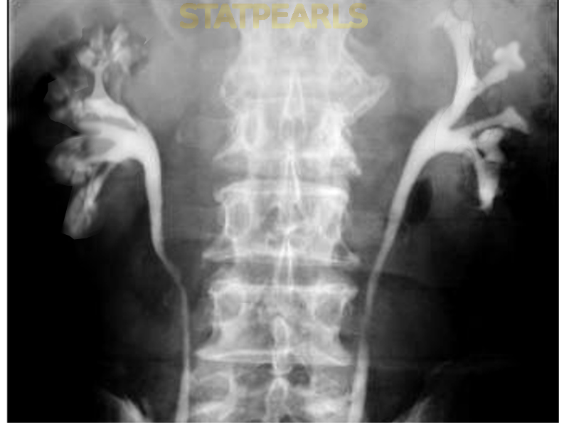
At intravenous urography (IVU), a classic paintbrush-like appearance within the dilated medullary collecting ducts is characteristic, but this test is no longer used in most clinical practices.
Multidetector contrast CT urography will show a distinctive papillary blush. On delayed imaging from a CT urogram, the characteristic finding of medullary sponge kidney is parallel striations from contrast which extend from the papilla to the medulla and persist on delayed imaging.
CT can also detect related complications such as hydronephrosis, calculi, and pyelonephritis. [16][17][18][19]
Treatment / Management
Treatment consists of managing the complications of medullary sponge kidney.
For UTIs, antibiotics and meticulous personal hygiene practices are recommended.
For calcium stones, initial recommendations include a high fluid intake sufficient to generate 2000 mL of urine per day. In general, a diet that is low in sodium, normal in calcium, high in potassium, and low to normal in protein may be helpful.
A 24-hour urine test is recommended to help optimize the urinary chemistry in motivated patients with medullary sponge kidney who develop stones. These patients will tend to have a higher incidence of renal leak type hypercalciuria and hypocitraturia than most calcium stone formers. If this is confirmed by the 24-hour urine test, these disorders can be treated with thiazide diuretics for the hypercalciuria and potassium citrate supplements for the hypocitraturia.
The potassium citrate supplementation also seems to help minimize the long-term bone loss that is sometimes associated with medullary sponge kidney. This bone loss is thought to be due primarily to the persistent renal leak type hypercalciuria although impaired urinary acidification has also been suggested. There is also a possible association with hyperparathyroidism.
Most of the stones in patients with medullary sponge kidney tend to be small and will usually pass spontaneously, but occasionally surgery, ureteroscopy, or lithotripsy may be needed. Overall, medullary sponge kidney patients who produce calcium stones tend to make about twice as many stones as other calcium stone formers.
Some patients will also have distal type renal tubular acidosis (RTA) which will demonstrate hypocitraturia and can then be treated with supplemental potassium citrate. The dosage of the potassium citrate should be titrated to approach an optimal 24-hour urinary citrate level (usually greater than 500 mg/24 hours) with a urinary pH around 6.5 if possible. A urinary pH over 7.2 to 7.5 should generally be avoided to minimize the production of calcium phosphate calculi. Serum potassium should also be monitored periodically to avoid hyperkalemia. [20][21][22][23]
Differential Diagnosis
The other causes of medullary nephrocalcinosis comprise the differential diagnosis of medullary sponge kidney. The differential diagnosis of medullary nephrocalcinosis is as follows: hyperparathyroidism, hypervitaminosis D, milk-alkali syndrome, and other pathological hypercalcemic or hypercalciuric states. Papillary necrosis can also cause medullary calcifications. A history of analgesic abuse will favor papillary necrosis. [24]
Prognosis
Although medullary sponge kidney is generally a benign condition, 10% of patients with medullary sponge kidney will ultimately develop renal failure. The majority of patients will have normal renal function throughout their lives. Renal failure is thought to arise from recurrent severe infections and extensive calculi formation. [25]
A small number of medullary sponge kidney patients will describe chronic, severe pain. This group tends to produce substantially more renal calculi than other MSK patients (3.1 stones per patient per year) and often require multiple hospitalizations for pain control. This suggests that 24-hour urine testing and aggressive metabolic treatment directed at nephrolithiasis prevention may be particularly worthwhile in this group of MSK patients. [2]
Complications
Recurrent nephrolithiasis (renal stones) is a major complication of medullary sponge kidney which often presents with renal colic, often accompanied by hematuria. Renal stones can result in UTI and urinary tract obstruction.
Pearls and Other Issues
Associations with the following disease entities have been reported:
- Ehlers-Danlos syndrome
- Marfan disease
- Congenital hemihypertrophy/Beckwith-Wiedemann syndrome (rare)
- Caroli disease
Although medullary sponge kidney is generally described as a benign condition, as many as 10% of patients will develop renal insufficiency or failure throughout their lives.
Since medullary sponge kidney is associated with hypercalciuria, especially the renal calcium leak type, those with the disease are at risk of developing osteopenia, osteoporosis and secondary hyperparathyroidism.
While there is no specific treatment for medullary sponge kidney, those with renal stones and hypercalciuria or UTIs should be treated appropriately for these conditions. [26][27]
Enhancing Healthcare Team Outcomes
Most patients with medullary sponge kidney should be seen at regular intervals by a urologist in addition to a primary care physician. [28]
Media
References
Gambaro G, Feltrin GP, Lupo A, Bonfante L, D'Angelo A, Antonello A. Medullary sponge kidney (Lenarduzzi-Cacchi-Ricci disease): a Padua Medical School discovery in the 1930s. Kidney international. 2006 Feb:69(4):663-70 [PubMed PMID: 16395272]
Xiang H, Han J, Ridley WE, Ridley LJ. Medullary sponge kidney. Journal of medical imaging and radiation oncology. 2018 Oct:62 Suppl 1():93-94. doi: 10.1111/1754-9485.40_12784. Epub [PubMed PMID: 30309197]
Vasudevan V, Samson P, Smith AD, Okeke Z. The genetic framework for development of nephrolithiasis. Asian journal of urology. 2017 Jan:4(1):18-26. doi: 10.1016/j.ajur.2016.11.003. Epub 2016 Nov 28 [PubMed PMID: 29264202]
Janjua MU,Long XD,Mo ZH,Dong CS,Jin P, Association of medullary sponge kidney and hyperparathyroidism with RET G691S/S904S polymorphism: a case report. Journal of medical case reports. 2018 Jul 9 [PubMed PMID: 29983117]
Level 3 (low-level) evidenceImam TH, Patail H, Patail H. Medullary Sponge Kidney: Current Perspectives. International journal of nephrology and renovascular disease. 2019:12():213-218. doi: 10.2147/IJNRD.S169336. Epub 2019 Sep 26 [PubMed PMID: 31576161]
Level 3 (low-level) evidenceFabris A, Anglani F, Lupo A, Gambaro G. Medullary sponge kidney: state of the art. Nephrology, dialysis, transplantation : official publication of the European Dialysis and Transplant Association - European Renal Association. 2013 May:28(5):1111-9. doi: 10.1093/ndt/gfs505. Epub 2012 Dec 9 [PubMed PMID: 23229933]
Level 3 (low-level) evidenceChatterjee R, Ramos E, Hoffman M, VanWinkle J, Martin DR, Davis TK, Hoshi M, Hmiel SP, Beck A, Hruska K, Coplen D, Liapis H, Mitra R, Druley T, Austin P, Jain S. Traditional and targeted exome sequencing reveals common, rare and novel functional deleterious variants in RET-signaling complex in a cohort of living US patients with urinary tract malformations. Human genetics. 2012 Nov:131(11):1725-38. doi: 10.1007/s00439-012-1181-3. Epub 2012 Jun 23 [PubMed PMID: 22729463]
Level 2 (mid-level) evidenceGambaro G,Danza FM,Fabris A, Medullary sponge kidney. Current opinion in nephrology and hypertension. 2013 Jul [PubMed PMID: 23680648]
Level 3 (low-level) evidenceSwonke ML, Mahmoud AM, Farran EJ, Dafashy TJ, Kerr PS, Kosarek CD, Sonstein J. Early Stone Manipulation in Urinary Tract Infection Associated with Obstructing Nephrolithiasis. Case reports in urology. 2018:2018():2303492. doi: 10.1155/2018/2303492. Epub 2018 Nov 25 [PubMed PMID: 30595937]
Level 3 (low-level) evidenceGeavlete P, Nita G, Alexandrescu E, Geavlete B. The impact of modern endourological techniques in the treatment of a century old disease--medullary sponge kidney with associated nephrolithiasis. Journal of medicine and life. 2013:6(4):482-5 [PubMed PMID: 24868267]
Level 2 (mid-level) evidenceMarien TP, Miller NL. Characteristics of renal papillae in kidney stone formers. Minerva urologica e nefrologica = The Italian journal of urology and nephrology. 2016 Dec:68(6):496-515 [PubMed PMID: 27441596]
Katabathina VS,Kota G,Dasyam AK,Shanbhogue AK,Prasad SR, Adult renal cystic disease: a genetic, biological, and developmental primer. Radiographics : a review publication of the Radiological Society of North America, Inc. 2010 Oct [PubMed PMID: 21071372]
Evan AP, Worcester EM, Williams JC Jr, Sommer AJ, Lingeman JE, Phillips CL, Coe FL. Biopsy proven medullary sponge kidney: clinical findings, histopathology, and role of osteogenesis in stone and plaque formation. Anatomical record (Hoboken, N.J. : 2007). 2015 May:298(5):865-77. doi: 10.1002/ar.23105. Epub 2015 Feb 17 [PubMed PMID: 25615853]
Marcelissen TA, Smits MA, Van Kerrebroeck P. [A woman with urinary tract infections and flank pain]. Nederlands tijdschrift voor geneeskunde. 2012:156(40):A3865 [PubMed PMID: 23031231]
Level 3 (low-level) evidenceMeyer A, Vetter W. [Recurrent flank pain, microhematuria]. Schweizerische Rundschau fur Medizin Praxis = Revue suisse de medecine Praxis. 1985 Nov 12:74(46):1286-8 [PubMed PMID: 4081442]
Level 3 (low-level) evidenceUchida T,Takechi H,Oshima N,Kumagai H, Medullary sponge kidney diagnosed by unenhanced magnetic resonance imaging. Iranian journal of kidney diseases. 2015 Jan [PubMed PMID: 25599731]
Level 3 (low-level) evidenceLeung JC. Inherited renal diseases. Current pediatric reviews. 2014:10(2):95-100 [PubMed PMID: 25088262]
Gaunay GS, Berkenblit RG, Tabib CH, Blitstein JR, Patel M, Hoenig DM. Efficacy of Multi-Detector Computed Tomography for the Diagnosis of Medullary Sponge Kidney. Current urology. 2018 Mar:11(3):139-143. doi: 10.1159/000447208. Epub 2018 Feb 20 [PubMed PMID: 29692693]
Koraishy FM, Ngo TT, Israel GM, Dahl NK. CT urography for the diagnosis of medullary sponge kidney. American journal of nephrology. 2014:39(2):165-70. doi: 10.1159/000358496. Epub 2014 Feb 11 [PubMed PMID: 24531190]
Level 3 (low-level) evidenceSun H,Zhang Z,Yuan J,Liu Y,Lei M,Luo J,Wan SP,Zeng G, Safety and efficacy of minimally invasive percutaneous nephrolithotomy in the treatment of patients with medullary sponge kidney. Urolithiasis. 2016 Oct [PubMed PMID: 26671346]
Bhojani N, Paonessa JE, Hameed TA, Worcester EM, Evan AP, Coe FL, Borofsky MS, Lingeman JE. Nephrocalcinosis in Calcium Stone Formers Who Do Not have Systemic Disease. The Journal of urology. 2015 Nov:194(5):1308-12. doi: 10.1016/j.juro.2015.05.074. Epub 2015 May 16 [PubMed PMID: 25988516]
Miano R, Germani S, Vespasiani G. Stones and urinary tract infections. Urologia internationalis. 2007:79 Suppl 1():32-6 [PubMed PMID: 17726350]
Carpentier X, Meria P, Bensalah K, Chabannes E, Estrade V, Denis E, Yonneau L, Mozer P, Hadjadj H, Hoznek A, Traxer O, Lithiasis Committee of the French Association of Urology. [Update for the management of kidney stones in 2013. Lithiasis Committee of the French Association of Urology]. Progres en urologie : journal de l'Association francaise d'urologie et de la Societe francaise d'urologie. 2014 Apr:24(5):319-26. doi: 10.1016/j.purol.2013.09.029. Epub 2013 Nov 22 [PubMed PMID: 24674339]
D'Alessandro MM, Pavone G, Castiglione MC, Tranchida AM, Sapia MC, Cusumano R, Corrado C, Mongiovì R, Maringhini S. [Nephrocalcinosis in children]. Giornale italiano di nefrologia : organo ufficiale della Societa italiana di nefrologia. 2015 Dec:35(Suppl 71):. pii: 2018-supp71. Epub [PubMed PMID: 29710439]
Cheungpasitporn W, Thongprayoon C, Brabec BA, Kittanamongkolchai W, Erickson SB. Outcomes of living kidney donors with medullary sponge kidney. Clinical kidney journal. 2016 Dec:9(6):866-870 [PubMed PMID: 27994868]
Adam MP, Feldman J, Mirzaa GM, Pagon RA, Wallace SE, Bean LJH, Gripp KW, Amemiya A, Shuman C, Kalish JM, Weksberg R. Beckwith-Wiedemann Syndrome. GeneReviews(®). 1993:(): [PubMed PMID: 20301568]
Chesney RW, Kaufman R, Stapleton FB, Rivas ML. Association of medullary sponge kidney and medullary dysplasia in Beckwith-Wiedemann syndrome. The Journal of pediatrics. 1989 Nov:115(5 Pt 1):761-4 [PubMed PMID: 2809912]
Level 3 (low-level) evidenceWikström B, Backman U, Danielson BG, Fellström B, Johansson G, Ljunghall S. Ambulatory diagnostic evaluation of 389 recurrent renal stone formers. A proposal for clinical classification and investigation. Klinische Wochenschrift. 1983 Jan 17:61(2):85-90 [PubMed PMID: 6843037]
