Introduction
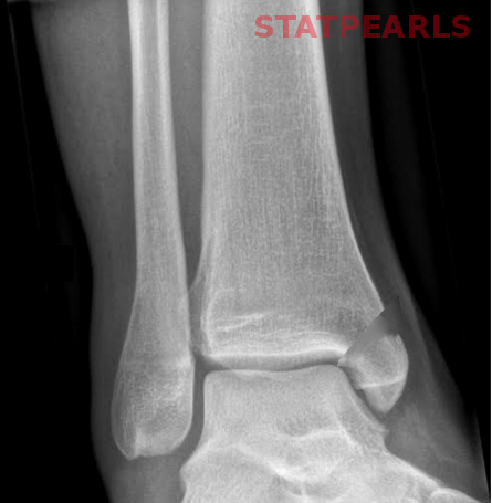
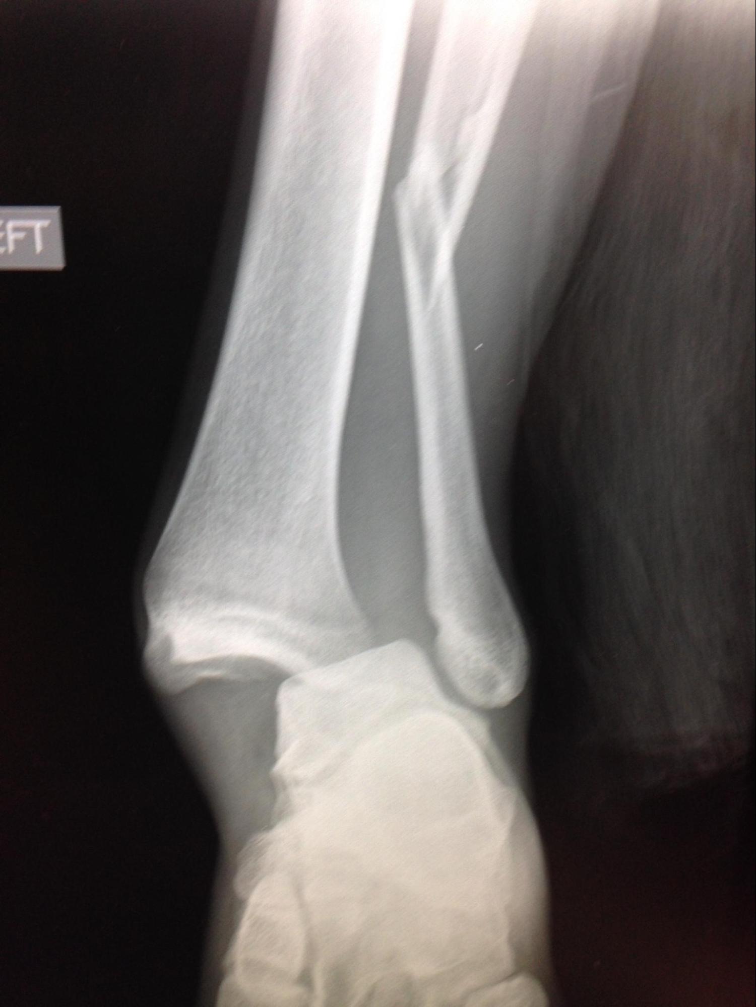
Growth plate or physeal fractures are common injuries in skeletally immature children and adolescents. Most fractures in skeletally immature individuals involve the physis as this cartilaginous growth center is the weakest part of the bone and, therefore, more susceptible to injury. Triplane ankle fractures are complex traumatic Salter-Harris IV fractures involving the metaphysis, physis, and epiphysis. The term “triplane” refers to the different orientations of the fracture lines in the distal tibia and represents a frequent diagnostic challenge. The epiphysis is fractured in the sagittal plane and is visible on the anteroposterior (AP) radiograph. The posterior aspect of the metaphysis is fractured in the coronal plane and appreciated on the lateral radiograph. The physis becomes separated in the axial plane. Treatment is closed reduction or surgical fixation depending on the degree of fracture displacement and articular step-off. The prognosis is excellent, given the triplane ankle fracture is identified and appropriately treated.
Etiology
Register For Free And Read The Full Article
Search engine and full access to all medical articles
10 free questions in your specialty
Free CME/CE Activities
Free daily question in your email
Save favorite articles to your dashboard
Emails offering discounts
Learn more about a Subscription to StatPearls Point-of-Care
Etiology
Triplane ankle fractures occur secondary to ankle trauma in an adolescent during a transitional period of partial physeal closure. Similar to Tillaux fractures (transitional Salter-Harris III ankle fracture), a supination-external rotational force is often the reported mechanism.[1] Lateral-sided triplane injuries are the most common as the lateral physis is the weakest and the point of insertion of the stout anterior inferior tibiofibular ligament (AITL).[2] Medial triplane injuries are rare and occur secondary to an adduction force.[3]
Epidemiology
Ankle injuries are common in children and rank second only to injuries to the hand and wrist in those aged 10 to 15 years.[4][5][6] Ankle fractures occur twice as frequently in males and represent 5% of all pediatric fractures and 9% to 18% of all physeal injuries.[3][5][7][8][9] Triplane fractures specifically account for 5% to 15% of pediatric ankle fractures and occur in adolescents with a mean age of 13 years and 5 months and a range of 10 to 17 years.[10] Tillaux fractures classically occur in older adolescents with more resulting physeal closure.[2]
Pathophysiology
Skeletal growth typically continues until 16 years in males and 14 years in females.[2][3] Physeal closure is driven by the hormone estrogen, which gets produced at a younger age in females. Physeal closure occurs in a predictable pattern beginning centrally and progresses anteromedially, posteromedially, and finally laterally.[2] Once closure of the distal tibia physis begins, there is an 18 to 20-month transitional period where closure is incomplete.[11] The unfused portions of the physis are at risk for transitional ankle fractures (triplane and Tillaux) during this period of incomplete growth plate closure. As the lateral aspect of the physis is the last to close, lateral epiphysis fractures are much more common than medial patterns.[2]
History and Physical
Adolescents typically report a twisting injury often during a sporting activity with resulting ankle pain and inability to bear weight. The most common injury mechanism is supination and external rotation, while the uncommon medial triplane fracture occurs with an adduction force.[1][3] Swelling or ecchymosis are commonly appreciated, but angular deformity can present in severe injuries. Gross instability is rarely appreciated. Patients will be tender to direct palpation of the physis circumferentially. As in all ankle injuries, the clinician should perform and document a thorough neurovascular exam.
Evaluation
Triplane ankle fractures are often under-appreciated with plain radiographs as each view typically only reveals a single fracture line. AP, mortise, and lateral views are essential. The AP view reveals the sagittal fracture line in the epiphysis (Salter-Harris III), and the lateral view demonstrating the coronal fracture line in the posterior metaphysis (Salter-Harris II). The mortise radiograph is the best way to appreciate articular displacement on plain films. Computed tomography (CT) is a vital tool for assessing the exact fracture pattern, degree of displacement, and articular step-off. Jones et al. demonstrated that all surgeons surveyed changed the starting point and trajectory of planned screw fixation after reviewing the CT scan versus relying on plain radiographs in a series of triplane ankle fractures.[12] Of note, the rare medial triplane fracture differs from the lateral injury in that the metaphyseal fracture occurs in the sagittal plane and the epiphysis injury is more medial and coronally oriented.
Treatment / Management
The treatment of triplane ankle fractures depends on the amount of fracture fragment displacement and degree of articular step-off visualized on CT. Nondisplaced and minimally displaced (less than 2 mm) injuries can effectively undergo management with long leg cast immobilization.[3] The reduction maneuver for the classic triplane ankle fracture pattern is ankle internal rotation with a post-reduction CT scan used to assess residual displacement and articular step-off. Rapariz et al. reported excellent outcomes in a series of triplane fractures displaced less than 2 mm, while displaced injuries developed chronic pain and ankle degenerative changes.[13]
Surgery is reserved for triplane fractures with over 2 mm of displacement or injuries that lost reduction during attempted nonoperative management. Fixation is typically achieved with one or two screws placed parallel to the physis. Screw placement can be in the metaphysis, epiphysis, or both depending on the fracture pattern. Screw types utilized for fixation vary widely no evidence suggesting the superiority of cannulated versus non-cannulated or fully threaded versus partially threaded screw fixation in triplane ankle fractures. Congruity of the articular surface must be restored to optimize outcomes.[11] Care is necessary to place screws perpendicularly as possible to fracture lines to maximize compression and maintain reduction. Clinicians and researchers have described both closed reduction and open reduction techniques with universally good outcomes.[14][15](B2)
Several clinicians' preferred technique includes percutaneous reduction via small incisions and fixation with one or two non-cannulated partially threaded 3.5 mm screws placed parallel to the physis. The epiphyseal fracture is usually amenable to anterolateral to posteromedial placed screws, while the metaphyseal fragment usually gets captured with direct anterior to posterior based screws. Care is also necessary to ensure all screw threads are past the fracture if using partially threaded screws. [Level 5]
Differential Diagnosis
Differential diagnoses of adolescent ankle pain include sprain, Tillaux fracture, triplane fracture, syndesmosis injury, ankle dislocation, subtalar dislocation, calcaneal fracture, talus fracture, malignancy, and infection.
Staging
Triplane ankle fractures are classified based on the number of parts as well as the pattern.
Classification by Parts
- 2-part
- Part 1: anterolateral and posterior epiphysis
- Part 2: anteromedial epiphysis
- 3-part
- Anterolateral epiphysis
- Posterior epiphysis
- Anteromedial epiphysis
- 4-part
- Comminuted
Classification by Pattern
- Lateral
- Most common
- Epiphyseal fracture: sagittal plane
- Physeal fracture: axial plane
- Metaphyseal fracture: coronal plane
- Medial
- Epiphyseal fracture: coronal plane
- Physeal fracture: axial plane
- Metaphyseal fracture: sagittal plane
- Intramalleolar
- Type I: intraarticular, involving weight-bearing surface
- Type II: intraarticular, not involving weight-bearing surface
- Type III: extraarticular
Prognosis
Appropriately identified and treated triplane ankle fractures have an excellent prognosis. Physeal damage or premature closure occurs in 7% to 21% of cases.[16][17][18][19] Many have questioned the significance of preserving the physis, given the limited remaining growth potential. The clinician should clinically follow patients with more than two years of expected growth should be followed clinically. Cooperman et al. reported a series of 14 triplane ankle fractures with three growth disturbance complications.[20] All occurred in patients with more than two years of growth remaining.[20]
Complications
Complications during nonoperative management include loss of reduction requiring operative fixation, nonunion, malunion, and persistent pain. Surgical adverse events are rare and include bleeding, infection, nonunion, painful hardware, and transient neuropathy.
Deterrence and Patient Education
A provider should not assume a pediatric ankle injury is simply a ligamentous sprain. The ligamentous stabilizers of the ankle are stronger than the physis making physeal fracture much more common. For appropriately aged patients, scrutinize orthogonal radiographic views in patients with apparent distal tibia Salter-Harris II or III fractures to avoid missing a more significant Salter-Harris IV triplane ankle fracture.
Enhancing Healthcare Team Outcomes
Treatment strategies of triplane ankle fractures depend on the residual displacement following reduction. Post-reduction CT scans should be obtained to assess the quality of reduction and articular step-off. Ertl et al. demonstrated that in a series of 23 triplane ankle fractures managed nonoperatively at a single institution, residual articular displacement of more than 2 mm results in poor outcomes if managed nonoperatively.[19]
Triplanar ankle fractures require an interprofessional team approach. The primary care practitioner should immediately enlist an orthopedist for evaluation and eventual treatment. Following any procedure or with non-operative management, an orthopedic specialty-trained nurse is invaluable. They can assist during surgery, provide post-surgical care, and help with conservative, non-operative management, monitor progress, and coordinate with physical therapy when necessary, serving as a coordination point between the treating clinician and other providers. This type of interprofessional collaboration will result in better outcomes and improved patient care. [Level 5]
Media
(Click Image to Enlarge)
References
Feldman DS, Otsuka NY, Hedden DM. Extra-articular triplane fracture of the distal tibial epiphysis. Journal of pediatric orthopedics. 1995 Jul-Aug:15(4):479-81 [PubMed PMID: 7560039]
Kay RM, Matthys GA. Pediatric ankle fractures: evaluation and treatment. The Journal of the American Academy of Orthopaedic Surgeons. 2001 Jul-Aug:9(4):268-78 [PubMed PMID: 11476537]
Wuerz TH, Gurd DP. Pediatric physeal ankle fracture. The Journal of the American Academy of Orthopaedic Surgeons. 2013 Apr:21(4):234-44. doi: 10.5435/JAAOS-21-04-234. Epub [PubMed PMID: 23545729]
King J, Diefendorf D, Apthorp J, Negrete VF, Carlson M. Analysis of 429 fractures in 189 battered children. Journal of pediatric orthopedics. 1988 Sep-Oct:8(5):585-9 [PubMed PMID: 3170740]
Peterson HA, Madhok R, Benson JT, Ilstrup DM, Melton LJ 3rd. Physeal fractures: Part 1. Epidemiology in Olmsted County, Minnesota, 1979-1988. Journal of pediatric orthopedics. 1994 Jul-Aug:14(4):423-30 [PubMed PMID: 8077422]
Level 2 (mid-level) evidenceMann DC, Rajmaira S. Distribution of physeal and nonphyseal fractures in 2,650 long-bone fractures in children aged 0-16 years. Journal of pediatric orthopedics. 1990 Nov-Dec:10(6):713-6 [PubMed PMID: 2250054]
Level 2 (mid-level) evidenceMizuta T, Benson WM, Foster BK, Paterson DC, Morris LL. Statistical analysis of the incidence of physeal injuries. Journal of pediatric orthopedics. 1987 Sep-Oct:7(5):518-23 [PubMed PMID: 3497947]
Level 2 (mid-level) evidencePeterson CA, Peterson HA. Analysis of the incidence of injuries to the epiphyseal growth plate. The Journal of trauma. 1972 Apr:12(4):275-81 [PubMed PMID: 5018408]
Worlock P, Stower M. Fracture patterns in Nottingham children. Journal of pediatric orthopedics. 1986 Nov-Dec:6(6):656-60 [PubMed PMID: 3793885]
Spiegel PG, Cooperman DR, Laros GS. Epiphyseal fractures of the distal ends of the tibia and fibula. A retrospective study of two hundred and thirty-seven cases in children. The Journal of bone and joint surgery. American volume. 1978 Dec:60(8):1046-50 [PubMed PMID: 721852]
Level 2 (mid-level) evidenceBlackburn EW, Aronsson DD, Rubright JH, Lisle JW. Ankle fractures in children. The Journal of bone and joint surgery. American volume. 2012 Jul 3:94(13):1234-44. doi: 10.2106/JBJS.K.00682. Epub [PubMed PMID: 22760393]
Jones S, Phillips N, Ali F, Fernandes JA, Flowers MJ, Smith TW. Triplane fractures of the distal tibia requiring open reduction and internal fixation. Pre-operative planning using computed tomography. Injury. 2003 May:34(4):293-8 [PubMed PMID: 12667783]
Rapariz JM, Ocete G, González-Herranz P, López-Mondejar JA, Domenech J, Burgos J, Amaya S. Distal tibial triplane fractures: long-term follow-up. Journal of pediatric orthopedics. 1996 Jan-Feb:16(1):113-8 [PubMed PMID: 8747367]
Lintecum N, Blasier RD. Direct reduction with indirect fixation of distal tibial physeal fractures: a report of a technique. Journal of pediatric orthopedics. 1996 Jan-Feb:16(1):107-12 [PubMed PMID: 8747366]
Level 2 (mid-level) evidenceCastellani C, Riedl G, Eberl R, Grechenig S, Weinberg AM. Transitional fractures of the distal tibia: a minimal access approach for osteosynthesis. The Journal of trauma. 2009 Dec:67(6):1371-5. doi: 10.1097/TA.0b013e31818866fd. Epub [PubMed PMID: 20009690]
Kärrholm J. The triplane fracture: four years of follow-up of 21 cases and review of the literature. Journal of pediatric orthopedics. Part B. 1997 Apr:6(2):91-102 [PubMed PMID: 9165437]
Level 1 (high-level) evidenceKling TF Jr, Bright RW, Hensinger RN. Distal tibial physeal fractures in children that may require open reduction. The Journal of bone and joint surgery. American volume. 1984 Jun:66(5):647-57 [PubMed PMID: 6725313]
El-Karef E, Sadek HI, Nairn DS, Aldam CH, Allen PW. Triplane fracture of the distal tibia. Injury. 2000 Nov:31(9):729-36 [PubMed PMID: 11084162]
Ertl JP, Barrack RL, Alexander AH, VanBuecken K. Triplane fracture of the distal tibial epiphysis. Long-term follow-up. The Journal of bone and joint surgery. American volume. 1988 Aug:70(7):967-76 [PubMed PMID: 3403587]
Cooperman DR, Spiegel PG, Laros GS. Tibial fractures involving the ankle in children. The so-called triplane epiphyseal fracture. The Journal of bone and joint surgery. American volume. 1978 Dec:60(8):1040-6 [PubMed PMID: 102648]

