Introduction
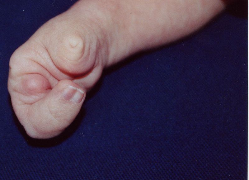
Cleft hand, otherwise referred to as ectrodactyly or colloquially as "split hand," is defined as a central longitudinal deficiency expressed as suppression of bone and soft tissues in the central elements of the hand, including the index, middle, and ring fingers.[1] Classically, this results in a "V-shaped" cleft in the hand with a variable degree of deformity. Generally, the phalanges of the affected digits are absent, and the metacarpals are present. The deficiency is typically bilateral.[2][3][4][5]
These central ray deformities typically divide into "typical" and "atypical" cleft hands. Typical cleft hands are generally of a genetic origin and bilateral with the classic "v-shaped" defect. Furthermore, typical cleft hands are often associated with "cleft foot" deformities as well. Atypical cleft hands, in contrast, are a form of symbrachydactyly involving the index, long, and ring fingers, often involving an absence of the digits, syndactyly of the digits, or hypoplasia of the digits. Atypical cleft hands generally appear as more of a "u-shaped" deformity compared to the classic "v-shape." Because atypical cleft hands are generally the result of a spontaneous mutation, there are rarely associated syndromes or deformities, and the disorder is not inherited.[2][6][7][4]
Etiology
Register For Free And Read The Full Article
Search engine and full access to all medical articles
10 free questions in your specialty
Free CME/CE Activities
Free daily question in your email
Save favorite articles to your dashboard
Emails offering discounts
Learn more about a Subscription to StatPearls Point-of-Care
Etiology
Typical cleft hand deformities are the result of an inherited autosomal dominant trait. The central rays develop differently from the thumb and small finger, and this mutation disrupts the central digit growth in the hands and often the feet. Atypical cleft hands, in contrast, are a result of a sporadic mutation and are therefore not inherited or associated with syndromic conditions.[2]
Epidemiology
The incidence of typical cleft hands is 1 in 90,000 births and 1 in 120,000 within the population. Atypical cleft hands occur in 1 in 150,000 births and 1 in 200,000 within the population. Bilateral cleft hands are observed in 56% of patients, while unilateral deformity is present in 44% of cases.[8]
History and Physical
Cleft hands are congenital disorders, and significant deformities are present at birth. An understanding of the exact deformity is critical to guide treatment. Family history for similar malformations will be suggestive of a typical cleft hand deformity, whereas an absence of family history will be more indicative of a sporadic mutation.
Clinicians should perform a careful assessment of the vasculature of the hand, ensuring the hand is well perfused as atypical cleft hands associated with Poland syndrome are a result of vascular hypoplasia. Furthermore, an examination of the ipsilateral chest wall will help to guide management. Absence or hypoplasia of the pectoralis musculature will be further evidence of vascular insufficiency at the subclavian artery and may necessitate staged surgical release of symbrachydactyly given the underlying poor vasculature.
Evaluation
Radiographs help guide the classification and surgical management of cleft hand deformities. Before undergoing any surgical treatment, surgeons must develop a plan that specifically addresses the patient's unique abnormality. In particular, surgeons must assess for the presence or absence of metacarpals in the central rays, the degree of symbrachydactyly that may be present, and especially the vascular supply to the hand. If the cleft hand is due to vascular insufficiency, as is the case in Poland syndrome, a staged surgical correction will be necessary.
Treatment / Management
While surgical management of a cleft foot is comparatively straightforward, surgical management of a cleft hand is variable and largely dependent on underlying anatomy. A small first web space between the thumb and index finger can be detrimental to the functional use of the hand, as the thumb is unable to have the same range of motion and lever arm present in an anatomically normal first webspace. Therefore, the functional goal of most surgeries is to split the cleft in half with an incision that wraps from the cleft and around the index finger into the first webspace. Often a flap from the ulnar half of the cleft is used to increase the depth of the first webspace, ultimately creating a wider and more functional space between the index finger and the thumb. Details of each surgical procedure are unique and based on each patient’s presenting deformity. Soft tissue releases, transfers, and osteotomies of the metacarpals may be necessary to achieve a more functional alignment.[9](B2)
Differential Diagnosis
Poland syndrome is attributed to subclavian hypoplasia leading to malformations of the limb during development. It is characterized by unilateral chest wall hypoplasia, symbrachydactyly, and shortening of the central fingers. When a unilateral atypical type cleft hand presents, clinicians should be aware of its association with Poland syndrome and evaluate for deficiency of the pectoralis musculature. In this scenario, patients who require syndactyly release should undergo surgery one digit at a time to avoid any vascular complications, as the underlying deficiency is due to vascular insufficiency during development.[10][11]
Prognosis
A cleft hand has been described as "a functional triumph and a social disaster," referring to the high degree of functionality of most hands with this deformity despite its atypical and malformed appearance.[3] Even so, surgical intervention, in most cases, improves the appearance of the hand without significantly affecting the functional outcome. In cases where the function is impaired, as when the first webspace is hypoplastic, surgical alignment of the hand often results in improved usage and cosmetic outcomes.[9]
In one study, researchers followed 23 patients with a cleft hand after having undergone surgical intervention to correct their deformities. Twenty-two of the 23 were satisfied with the aesthetics of the hand after surgical intervention, and 21 of the 23 reported being "satisfied" or "very satisfied" with their functional outcome. The most common complaint after surgery is digital stiffness.[3]
Complications
Patients who have a cleft hand due to a vascular deformity are at high risk for skin loss and poor perfusion of the surgical site after surgery, particularly if the procedure does not have appropriate staging. Furthermore, stiffness of the digits remains the most common postoperative complaint despite improved overall functional outcomes.
Deterrence and Patient Education
Parents of children with cleft hand deformities should seek treatment from a pediatric orthopedic specialist to determine the most appropriate treatment. Surgical intervention has cosmetic benefits, and in some cases, a functional benefit. Correction may be difficult and should be individualized for each patient, but children with a cleft hand deformity frequently lead normal lives with little or no functional limitations when managed appropriately.[9]
Enhancing Healthcare Team Outcomes
Cleft hand deformities are rare and therefore require the intervention of a pediatric orthopedic specialist who can adequately address the deformity to maximize function and aesthetics as well as to assess the patient for additional developmental abnormalities properly. To have an optimal outcome, the surgical and nursing staff must work together to execute the plan of management appropriately. Finally, members of the interdisciplinary team, which may include pediatric counselors for age-appropriate patients, must communicate clearly with the patient and family to help them understand and overcome the challenges that present with cleft hand deformities. [Level V]
Media
(Click Image to Enlarge)
References
Sharma A, Sharma N. A comprehensive functional classification of cleft hand: The DAST concept. Indian journal of plastic surgery : official publication of the Association of Plastic Surgeons of India. 2017 Sep-Dec:50(3):244-250. doi: 10.4103/ijps.IJPS_8_17. Epub [PubMed PMID: 29618858]
Kozin SH. Upper-extremity congenital anomalies. The Journal of bone and joint surgery. American volume. 2003 Aug:85(8):1564-76 [PubMed PMID: 12925640]
Level 3 (low-level) evidenceAleem AW, Wall LB, Manske MC, Calhoun V, Goldfarb CA. The transverse bone in cleft hand: a case cohort analysis of outcome after surgical reconstruction. The Journal of hand surgery. 2014 Feb:39(2):226-36. doi: 10.1016/j.jhsa.2013.11.002. Epub 2013 Dec 18 [PubMed PMID: 24359797]
Level 2 (mid-level) evidenceManske PR,Halikis MN, Surgical classification of central deficiency according to the thumb web. The Journal of hand surgery. 1995 Jul; [PubMed PMID: 7594304]
Goldfarb CA, Chia B, Manske PR. Central ray deficiency: subjective and objective outcome of cleft reconstruction. The Journal of hand surgery. 2008 Nov:33(9):1579-88. doi: 10.1016/j.jhsa.2008.05.010. Epub [PubMed PMID: 18984341]
Level 2 (mid-level) evidenceBARSKY AJ. CLEFT HAND: CLASSIFICATION, INCIDENCE, AND TREATMENT. REVIEW OF THE LITERATURE AND REPORT OF NINETEEN CASES. The Journal of bone and joint surgery. American volume. 1964 Dec:46():1707-20 [PubMed PMID: 14239859]
Level 3 (low-level) evidenceOgino T. Teratogenic relationship between polydactyly, syndactyly and cleft hand. Journal of hand surgery (Edinburgh, Scotland). 1990 May:15(2):201-9 [PubMed PMID: 2164076]
Level 3 (low-level) evidenceBaba AN, Bhat YJ, Ahmed SM, Nazir A. Unilateral cleft hand with cleft foot. International journal of health sciences. 2009 Jul:3(2):243-6 [PubMed PMID: 21475543]
Level 3 (low-level) evidenceUpton J. Simplicity and treatment of the typical cleft hand. Handchirurgie, Mikrochirurgie, plastische Chirurgie : Organ der Deutschsprachigen Arbeitsgemeinschaft fur Handchirurgie : Organ der Deutschsprachigen Arbeitsgemeinschaft fur Mikrochirurgie der Peripheren Nerven und Gefasse : Organ der V.... 2004 Apr-Jun:36(2-3):152-60 [PubMed PMID: 15162314]
Level 2 (mid-level) evidenceIreland DC, Takayama N, Flatt AE. Poland's syndrome. The Journal of bone and joint surgery. American volume. 1976 Jan:58(1):52-8 [PubMed PMID: 175070]
Level 3 (low-level) evidenceCatena N, Divizia MT, Calevo MG, Baban A, Torre M, Ravazzolo R, Lerone M, Sénès FM. Hand and upper limb anomalies in Poland syndrome: a new proposal of classification. Journal of pediatric orthopedics. 2012 Oct-Nov:32(7):727-31. doi: 10.1097/BPO.0b013e318269c898. Epub [PubMed PMID: 22955538]
Level 2 (mid-level) evidence