Introduction
Radiation therapy is an important component in the treatment of cancer. It may play a role as an adjuvant, neoadjuvant, palliative, or definitive therapy, with or without concurrent chemotherapy. Radiation is most commonly delivered as a local/regional treatment by an external beam consisting of photons, electrons, protons, or heavy particles but may also be delivered via brachytherapy (where a sealed radiation source is placed adjacent to the target) or systemically via unsealed sources. The side effects of radiation therapy are a function of the tissues included in the radiation field. Treatment of diseases within the thoracic region, such as Hodgkin lymphoma, lung, and breast cancer, carries the risk of radiation-induced cardiovascular toxicity (RICT).[1]
Side effects of therapeutic radiation to the heart and coronary vessels include pericarditis, coronary artery disease (CAD), arrhythmias, cardiomyopathy, valvular dysfunction, and heart failure. Pericarditis and pericardial effusions are potential short-term toxicities that may occur during or within the weeks following treatment. Long-term side effects may present in the months to years after radiation therapy, possibly as late as 20 years or more post-treatment. Late toxicities include CAD, valvular heart disease, and heart failure.[1][2]
Major risk factors that increase the likelihood of RICT include higher radiation doses, adjuvant treatment with cardiotoxic chemotherapy, irradiation of the left side of the thorax, and the presence of pre-existing cardiovascular disease.[3][4] Studies have correlated the mean dose of radiation received by various heart sub-structures to the incidence of major adverse cardiac events (MACE), such as hospitalization for heart failure, myocardial infarction, and even cardiac death.[5] Given the importance of radiation therapy in treating cancer and the high prevalence of cardiovascular disease in Western populations, numerous preventive measures have been suggested and used in clinical practice, such as dose limitation, proton and particle therapy, conformal radiation therapy, and deep-inspiration breath-hold technique.[3][6]
Issues of Concern
Register For Free And Read The Full Article
Search engine and full access to all medical articles
10 free questions in your specialty
Free CME/CE Activities
Free daily question in your email
Save favorite articles to your dashboard
Emails offering discounts
Learn more about a Subscription to StatPearls Point-of-Care
Issues of Concern
When any tissue is irradiated, the cells comprising the tissue are damaged primarily by the generation of free radicals, predominantly by the hydroxyl radical. In addition to inducing pro-inflammatory cytokines, free radicals react with DNA to cause strand disruption, preventing proper replication and protein synthesis. Cardiomyocytes are stable cells and are therefore relatively resistant to radiation. However, modern therapeutic doses of radiation are sufficient to cause damage to the microvasculature of the myocardium.[7][8] Damage to the microvasculature results in structural changes of the heart through pericardial inflammation, fibrosis of the myocardium, valvular dysfunction, fibrosis of the electrical conduction system, and endothelial damage in the coronary vessels.
The pericardium may become acutely inflamed, leading to pericarditis or pericardial effusion. The pericardium can also develop chronic fibrosis, resulting in constrictive pericarditis. In the myocardium, the diffuse development of infiltrative fibrosis impairs the ability of the ventricles to relax, resulting in diastolic failure. These fibrotic changes can also affect the conduction system in the heart and result in the development of arrhythmias later in life. In the years following radiation therapy, the valvular endothelium can break down or become fibrotic, leading to regurgitation or stenosis.[9] The pro-inflammatory effect of radiation on the coronary vasculature mimics atherosclerosis in that endothelial damage promotes fibrin deposition and intimal proliferation, thereby hastening the progression of coronary artery disease.[7]
Clinical Significance
Specific radiation doses are communicated in Gray (Gy), a measure of absorbed radiation that is the predominant unit used in clinical practice, and the Sievert (Sv), which considers the relative biological effectiveness of the specific type of radiation within the tissue. Both units measure one joule of energy absorbed per kilogram. RICT is both a short and long-term concern, with the median time to diagnosis estimated to be 19 years.[1] The heart is affected by radiation in a dose-dependent manner with higher radiation doses, particularly >40 Gy, associated with significantly increased post-radiation-induced mortality.[1] However, damaging effects can also be seen after doses as low as 2 Gy, and there is no apparent "safe" dose of radiation that the heart may receive.[10][11] Further risk factors associated with radiation-induced heart disease include earlier age at treatment, irradiation of left-sided cancers, concurrent treatment with trastuzumab or anthracyclines, pre-existing cardiovascular disease, and the presence of general cardiovascular risk factors (such as diabetes and hypertension).[11][12][13] RICT may affect any sub-system of the heart and therefore has a varied presentation. In the acute setting, pericarditis and pericardial effusions may manifest during or within the initial weeks following irradiation and may be treated similarly to acute pericarditis or effusion unrelated to radiation. Delayed pericarditis, characterized by thickening and fibrosis of the pericardium, can result in restrictive pericarditis and subsequent diastolic heart dysfunction. Diseases of the pericardium are more common at doses >40 Gy.[14][15]
Valvular disease is a late complication. Fibrosis, calcification, and thickening of the valves can occur asymptomatically for over 15 years before becoming clinically apparent. Prominent damage is more common in the left-sided valves and can manifest as stenosis, regurgitation, or a proclivity towards endocarditis. Valvular effects can occur at doses >30 Gy.[13][15][16] The myocardium itself is affected by the progressive loss of capillary beds due to oxidative stress, DNA damage, and microvasculature inflammation.[17] This damage results in diffuse fibrosis of the myocardium, leading to stiffening, impaired ventricular filling, and potentially leading to restrictive cardiomyopathy (i.e., diastolic heart failure), and eventually systolic heart failure. Severe cardiomyopathy is more prevalent when radiation therapy is combined with anthracyclines, such as daunorubicin and doxorubicin, and the monoclonal antibody trastuzumab - though this effect appears to be additive as opposed to synergistic.[11] Cardiomyocyte toxicity and cardiomyopathy are most common in radiation doses >30 Gy.[14][15]
Arrhythmias are another late effect of radiation therapy due to toxicity to the sinoatrial (SA) and atrioventricular (AV) nodes and the conduction system of the heart. Transient and asymptomatic arrhythmia may occur within a year of therapy, but permanent damage to the cardiac nodes and bundle branch blocks may manifest ten or more years after treatment completion. The association between the development of arrhythmias and the dose received has not been thoroughly studied; therefore, the absorbed dose associated with arrhythmias is not well described.[13][15]
Atherosclerosis is worsened by radiation therapy, as the inflammatory effects of radiation accelerate the damage to the vascular endothelium. Therefore, premature or worsened coronary artery disease (CAD) is another potential complication of radiation therapy to the heart and increases the risk of unstable angina, myocardial infarction, and cardiac mortality.[10][17] An independent association has been shown between the dose received by the left anterior descending artery (LAD) and MACE, including myocardial infarction (MI) and hospitalization for heart failure.[5] Additionally, pre-existing CAD and atherosclerosis affect the dose that the left ventricle may receive. Patients with traditional CAD risk factors such as diabetes mellitus, smoking, hypertension, hyperlipidemia, and male gender are more likely to experience MACE after thoracic radiation therapy.[12][15][5] Atherosclerotic heart disease can be exacerbated by radiation doses as low as 6 Gy.[14] A linear association has been shown between mean cardiac dose and RICT, with a 7.4% relative increase in rates of coronary events with each additional Gray, without an observed threshold.[11]
Given the potentially serious consequences of cardiac damage during radiation therapy, techniques for mitigating collateral cardiac irradiation have been developed. One option is using proton or heavy particle (e.g., carbon ion) therapy due to the theoretical advantage offered by reducing the exit dose characteristic to such particles. Treatment in the prone position is an option for breast cancer that allows the breast to displace through an aperture in the treatment table away from the body that has shown to reduce heart and lung dose received over traditional supine treatment.[18]
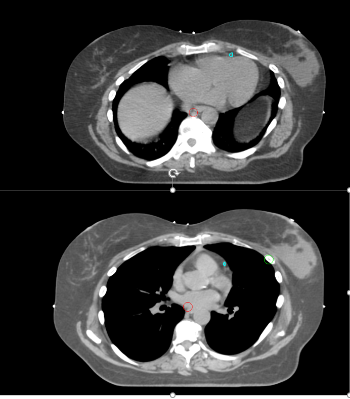
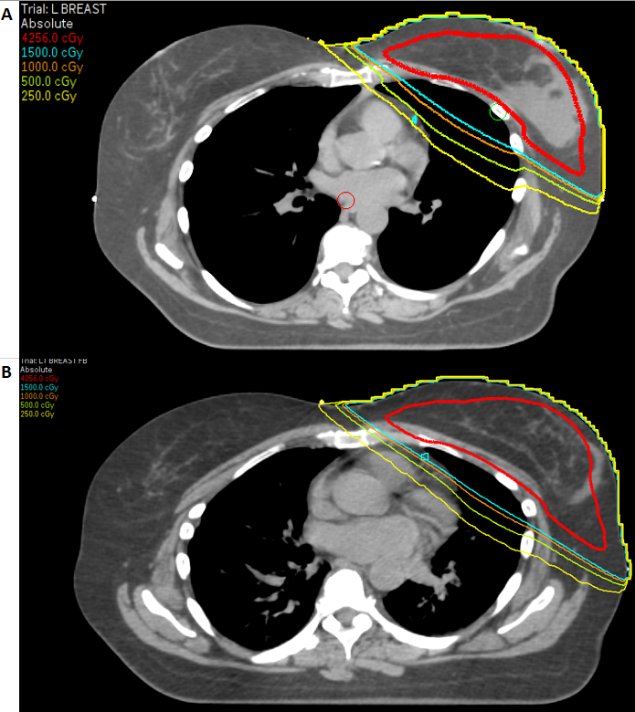
A promising option for the reduction of heart dose in patients with left-sided breast cancer is the deep inspiration breath-hold (DIBH) technique, in which a patient is asked to hold a moderately deep breath while lying supine to increase lung filling, thus moving the chest wall target further from the heart [see attached image]. Surface imaging is used to measure chest rise at the time of simulation and treatment, and treatment is only delivered while the lungs are expanded into the appropriate position, allowing for reproducibility during each fraction and assurance of treatment delivery as planned. Multiple studies have shown up to a 40-50% relative reduction in dose received by the heart as a result of this technique, with mean LAD doses of 2-3Gy.[10][14][19]
In treating lung and esophageal cancer and Hodgkin lymphoma, or when DIBH is not available for left breast cancer, highly conformal therapies such as intensity-modulated radiation therapy (IMRT) may be used to minimize radiation dose to the heart as compared with older 3D planning techniques.[14][20][21] This is done by utilizing multiple non-uniform energy fields in such a way that the high dose region conforms tightly to the target but decreases quickly in the surrounding normal tissues. IMRT has been shown by a prospective NRG Oncology study focusing on lung cancer to aid in reducing the heart V40 from 11.4% to 6.8%, as compared with conventional 3D planning.[20][21] IMRT has also been shown to significantly reduce the dose received by the heart in whole lung irradiation, with 99% of the dose received by the heart when using a 3D plan vs. only 33% of the dose when an IMRT plan is utilized.[22]
It is important to identify pre-existing risk factors for heart disease in cancer patients before administering thoracic radiation therapy, with relevant intervention including smoking cessation counseling, as well as ensuring adequate treatment of hypertension, hyperlipidemia, and diabetes. Additionally, a full cardiac evaluation including EKG and echocardiogram should be performed prior to radiation therapy when planned to be combined with anthracyclines or trastuzumab.[23] When radiation therapy is indicated, patients with impaired cardiac function at baseline may be considered for the omission of systemic therapy in a risk vs. benefit analysis on a case-to-case basis, acknowledging that omitting any therapy places the patient at increased risk for sub-optimal oncologic outcomes.
There are currently no official screening guidelines for radiation-induced cardiac toxicity. However, given the known interplay of radiation with pre-existing cardiovascular risk factors, it is reasonable to suggest that identifying these at-risk patients and close follow-up for all could facilitate earlier diagnosis and treatment of any radiation-induced cardiac toxicities. When evaluating potential heart disease, imaging including cardiac MRI, CT, echocardiography, and myocardial perfusion studies are high-yield diagnostic tests that may guide further intervention.[16][17] However, cardiac MRI is considered the best investigatory option for evaluating radiation-induced heart disease, given that it aids in understanding the pathology and severity of the specific radiation-induced heart toxicity.[12][15]
Enhancing Healthcare Team Outcomes
Generally, the exact detail and pathophysiology of radiation-induced cardiac toxicity are beyond the scope of this portion of the discussion. There are no official published screening or prevention guidelines. Therefore, no precise recommendations are possible. However, there are risks associated with worsened cardiac outcomes following radiation therapy. These include younger age at treatment, radiation doses above 30 Gy (although there is no truly safe dose of radiation), irradiation of left-sided breast cancers or intrathoracic lesions, pre-existing heart disease, and the presence of risk factors for coronary artery disease: diabetes, hypertension, smoking, and hyperlipidemia. The interprofessional team must identify these patients before receiving radiation therapy to optimize lifestyle and pharmacologic interventions and coordinate proper follow-up and care. Nursing and other medical support staff play a critical role in identifying these patients and ensuring that the providing physician is aware of these risk factors to discuss extended cardiac follow-up with the patient. Additionally, nurses involved in the patient intake should remain vigilant for any patient with a remote history of thoracic radiation therapy showing signs or symptoms suggestive of cardiovascular disease. Appropriate identification of risk factors, adequate patient follow-up, and anticipation of cardiac complications are key factors to significantly improve the morbidity and mortality in post-radiation cancer patients.[10][15] [Level 5]
Media
(Click Image to Enlarge)
(Click Image to Enlarge)
References
Lenneman CG, Sawyer DB. Cardio-Oncology: An Update on Cardiotoxicity of Cancer-Related Treatment. Circulation research. 2016 Mar 18:118(6):1008-20. doi: 10.1161/CIRCRESAHA.115.303633. Epub [PubMed PMID: 26987914]
Curigliano G, Cardinale D, Dent S, Criscitiello C, Aseyev O, Lenihan D, Cipolla CM. Cardiotoxicity of anticancer treatments: Epidemiology, detection, and management. CA: a cancer journal for clinicians. 2016 Jul:66(4):309-25. doi: 10.3322/caac.21341. Epub 2016 Feb 26 [PubMed PMID: 26919165]
Rygiel K. Cardiotoxic effects of radiotherapy and strategies to reduce them in patients with breast cancer: An overview. Journal of cancer research and therapeutics. 2017 Apr-Jun:13(2):186-192. doi: 10.4103/0973-1482.187303. Epub [PubMed PMID: 28643731]
Level 3 (low-level) evidenceNiska JR, Thorpe CS, Allen SM, Daniels TB, Rule WG, Schild SE, Vargas CE, Mookadam F. Radiation and the heart: systematic review of dosimetry and cardiac endpoints. Expert review of cardiovascular therapy. 2018 Dec:16(12):931-950. doi: 10.1080/14779072.2018.1538785. Epub 2018 Nov 1 [PubMed PMID: 30360659]
Level 1 (high-level) evidenceAtkins KM, Chaunzwa TL, Lamba N, Bitterman DS, Rawal B, Bredfeldt J, Williams CL, Kozono DE, Baldini EH, Nohria A, Hoffmann U, Aerts HJWL, Mak RH. Association of Left Anterior Descending Coronary Artery Radiation Dose With Major Adverse Cardiac Events and Mortality in Patients With Non-Small Cell Lung Cancer. JAMA oncology. 2021 Feb 1:7(2):206-219. doi: 10.1001/jamaoncol.2020.6332. Epub [PubMed PMID: 33331883]
Bergom C, Currey A, Desai N, Tai A, Strauss JB. Deep Inspiration Breath Hold: Techniques and Advantages for Cardiac Sparing During Breast Cancer Irradiation. Frontiers in oncology. 2018:8():87. doi: 10.3389/fonc.2018.00087. Epub 2018 Apr 4 [PubMed PMID: 29670854]
Halle M, Gabrielsen A, Paulsson-Berne G, Gahm C, Agardh HE, Farnebo F, Tornvall P. Sustained inflammation due to nuclear factor-kappa B activation in irradiated human arteries. Journal of the American College of Cardiology. 2010 Mar 23:55(12):1227-1236. doi: 10.1016/j.jacc.2009.10.047. Epub [PubMed PMID: 20298930]
Hooning MJ, Aleman BM, van Rosmalen AJ, Kuenen MA, Klijn JG, van Leeuwen FE. Cause-specific mortality in long-term survivors of breast cancer: A 25-year follow-up study. International journal of radiation oncology, biology, physics. 2006 Mar 15:64(4):1081-91 [PubMed PMID: 16446057]
Orzan F, Brusca A, Gaita F, Giustetto C, Figliomeni MC, Libero L. Associated cardiac lesions in patients with radiation-induced complete heart block. International journal of cardiology. 1993 May:39(2):151-6 [PubMed PMID: 8314649]
Yeboa DN, Evans SB. Contemporary Breast Radiotherapy and Cardiac Toxicity. Seminars in radiation oncology. 2016 Jan:26(1):71-8. doi: 10.1016/j.semradonc.2015.09.003. Epub 2015 Sep 4 [PubMed PMID: 26617212]
Darby SC, Ewertz M, McGale P, Bennet AM, Blom-Goldman U, Brønnum D, Correa C, Cutter D, Gagliardi G, Gigante B, Jensen MB, Nisbet A, Peto R, Rahimi K, Taylor C, Hall P. Risk of ischemic heart disease in women after radiotherapy for breast cancer. The New England journal of medicine. 2013 Mar 14:368(11):987-98. doi: 10.1056/NEJMoa1209825. Epub [PubMed PMID: 23484825]
Level 2 (mid-level) evidenceWalker CM, Saldaña DA, Gladish GW, Dicks DL, Kicska G, Mitsumori LM, Reddy GP. Cardiac complications of oncologic therapy. Radiographics : a review publication of the Radiological Society of North America, Inc. 2013 Oct:33(6):1801-15. doi: 10.1148/rg.336125005. Epub [PubMed PMID: 24108563]
Adams MJ, Lipsitz SR, Colan SD, Tarbell NJ, Treves ST, Diller L, Greenbaum N, Mauch P, Lipshultz SE. Cardiovascular status in long-term survivors of Hodgkin's disease treated with chest radiotherapy. Journal of clinical oncology : official journal of the American Society of Clinical Oncology. 2004 Aug 1:22(15):3139-48 [PubMed PMID: 15284266]
Menezes KM, Wang H, Hada M, Saganti PB. Radiation Matters of the Heart: A Mini Review. Frontiers in cardiovascular medicine. 2018:5():83. doi: 10.3389/fcvm.2018.00083. Epub 2018 Jul 9 [PubMed PMID: 30038908]
Lancellotti P, Nkomo VT, Badano LP, Bergler-Klein J, Bogaert J, Davin L, Cosyns B, Coucke P, Dulgheru R, Edvardsen T, Gaemperli O, Galderisi M, Griffin B, Heidenreich PA, Nieman K, Plana JC, Port SC, Scherrer-Crosbie M, Schwartz RG, Sebag IA, Voigt JU, Wann S, Yang PC, European Society of Cardiology Working Groups on Nuclear Cardiology and Cardiac Computed Tomography and Cardiovascular Magnetic Resonance, American Society of Nuclear Cardiology, Society for Cardiovascular Magnetic Resonance, Society of Cardiovascular Computed Tomography. Expert consensus for multi-modality imaging evaluation of cardiovascular complications of radiotherapy in adults: a report from the European Association of Cardiovascular Imaging and the American Society of Echocardiography. European heart journal. Cardiovascular Imaging. 2013 Aug:14(8):721-40. doi: 10.1093/ehjci/jet123. Epub [PubMed PMID: 23847385]
Level 3 (low-level) evidenceHeidenreich PA, Hancock SL, Lee BK, Mariscal CS, Schnittger I. Asymptomatic cardiac disease following mediastinal irradiation. Journal of the American College of Cardiology. 2003 Aug 20:42(4):743-9 [PubMed PMID: 12932613]
Domercant J, Polin N, Jahangir E. Cardio-Oncology: A Focused Review of Anthracycline-, Human Epidermal Growth Factor Receptor 2 Inhibitor-, and Radiation-Induced Cardiotoxicity and Management. The Ochsner journal. 2016 Fall:16(3):250-6 [PubMed PMID: 27660573]
Formenti SC, Gidea-Addeo D, Goldberg JD, Roses DF, Guth A, Rosenstein BS, DeWyngaert KJ. Phase I-II trial of prone accelerated intensity modulated radiation therapy to the breast to optimally spare normal tissue. Journal of clinical oncology : official journal of the American Society of Clinical Oncology. 2007 Jun 1:25(16):2236-42 [PubMed PMID: 17470849]
Swanson T, Grills IS, Ye H, Entwistle A, Teahan M, Letts N, Yan D, Duquette J, Vicini FA. Six-year experience routinely using moderate deep inspiration breath-hold for the reduction of cardiac dose in left-sided breast irradiation for patients with early-stage or locally advanced breast cancer. American journal of clinical oncology. 2013 Feb:36(1):24-30. doi: 10.1097/COC.0b013e31823fe481. Epub [PubMed PMID: 22270108]
Boyle J, Ackerson B, Gu L, Kelsey CR. Dosimetric advantages of intensity modulated radiation therapy in locally advanced lung cancer. Advances in radiation oncology. 2017 Jan-Mar:2(1):6-11. doi: 10.1016/j.adro.2016.12.006. Epub 2017 Jan 3 [PubMed PMID: 28740910]
Level 3 (low-level) evidenceChun SG, Hu C, Choy H, Komaki RU, Timmerman RD, Schild SE, Bogart JA, Dobelbower MC, Bosch W, Galvin JM, Kavadi VS, Narayan S, Iyengar P, Robinson CG, Wynn RB, Raben A, Augspurger ME, MacRae RM, Paulus R, Bradley JD. Impact of Intensity-Modulated Radiation Therapy Technique for Locally Advanced Non-Small-Cell Lung Cancer: A Secondary Analysis of the NRG Oncology RTOG 0617 Randomized Clinical Trial. Journal of clinical oncology : official journal of the American Society of Clinical Oncology. 2017 Jan:35(1):56-62 [PubMed PMID: 28034064]
Level 1 (high-level) evidenceKalapurakal JA, Gopalakrishnan M, Walterhouse DO, Rigsby CK, Rademaker A, Helenowski I, Kessel S, Morano K, Laurie F, Ulin K, Esiashvili N, Katzenstein H, Marcus K, Followill DS, Wolden SL, Mahajan A, Fitzgerald TJ. Cardiac-Sparing Whole Lung IMRT in Patients With Pediatric Tumors and Lung Metastasis: Final Report of a Prospective Multicenter Clinical Trial. International journal of radiation oncology, biology, physics. 2019 Jan 1:103(1):28-37. doi: 10.1016/j.ijrobp.2018.08.034. Epub 2018 Aug 29 [PubMed PMID: 30170102]
Level 1 (high-level) evidenceVolkova M, Russell R 3rd. Anthracycline cardiotoxicity: prevalence, pathogenesis and treatment. Current cardiology reviews. 2011 Nov:7(4):214-20 [PubMed PMID: 22758622]

